DNA packaging & counting 1. Cell Cycle
G₁ → S → G₂ → M 2. Mitosis
copy → align → separate 3. Meiosis
halve + diversify 4. Compare
mitosis vs meiosis 5. Clinical
cancer & aneuploidy 6. Reproduction
gametes → zygote 7. Replication
copy DNA accurately 8. Central Dogma
DNA → RNA → protein 9. Regulation
turn genes on/off
🧬 0. Introduction: Genomes, Chromatin, and Chromosomes
The ability of organisms to reproduce their own kind is the one characteristic that best distinguishes living things from nonliving matter. The continuity of life is based entirely on the reproduction of cells, or Cell Division. Before a cell can divide, it must flawlessly organize, duplicate, and segregate its massive library of genetic information.
The Organization of Eukaryotic DNA
- Genome: A cell's total endowment of DNA. In prokaryotes, it is typically a single circular DNA molecule located in the nucleoid region. In eukaryotes, the genome is vastly larger and consists of multiple discrete linear DNA molecules enclosed within a double-membraned nucleus.
- Chromatin: Eukaryotic chromosomes are composed of chromatin, a highly complex, dynamic structural array of DNA intimately associated with specific proteins. During interphase (non-dividing states), chromatin exists as a diffuse, tangled mass.
- Euchromatin: Loosely packed, lightly staining DNA. This region is transcriptionally active, meaning RNA polymerase can access the genes to transcribe mRNA.
- Heterochromatin: Tightly packed, heavily staining, highly condensed DNA. This region is transcriptionally inactive. Centromeres and telomeres are largely composed of constitutive heterochromatin.
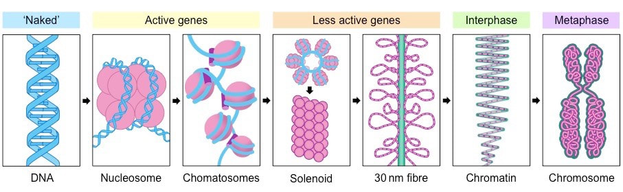
- Histones & Nucleosomes: To fit roughly 2 meters of human DNA into a nucleus that is only about $10 \mu m$ in diameter, it must be massively compacted. The DNA double helix wraps exactly twice around a core octamer of 8 basic histone proteins (two each of H2A, H2B, H3, H4) to form a Nucleosome (often described as "beads on a string"). A fifth histone protein (H1) sits outside the core and clamps the DNA to the nucleosome, facilitating higher-order packing into the 30-nm fiber.
- Chromosomes: Strictly prior to cell division, the chromatin supercoils, folds, and condenses massively to form distinct, heavily visible bodies called chromosomes. This extreme physical condensation prevents the delicate, incredibly long DNA strands from tangling, knotting, and breaking during the violent mechanical movements of cellular division.
Somatic cells (all general body cells except the gametes) contain two full sets of chromosomes, one inherited from each parent, making them Diploid (2n). In humans, the diploid number is 2n = 46 (comprising 22 pairs of autosomes and 1 pair of sex chromosomes). Gametes (sperm and eggs) contain only a single, unique set of chromosomes, making them Haploid (n). In humans, n = 23.
Figure 0.1A: Nucleosome core complex wrapping DNA around histone octamers ("beads on a string" 11nm fiber).
Figure 0.1B: Hierarchical packaging from solenoid 30nm fiber to looped radial domains and fully condensed metaphase chromosomes.

Anatomy of a Duplicated Chromosome
Following DNA replication during the S phase, each chromosome consists of two absolutely identical copies. These are formally called Sister Chromatids. They are initially attached firmly together all along their lengths by ring-like protein complexes called Cohesins. They are most tightly and visibly bound at a highly constricted region of DNA called the Centromere. Once the sister chromatids separate during anaphase of mitosis (or anaphase II of meiosis), they are no longer considered chromatids; they are immediately considered full, individual, independent chromosomes.

| Human cell state | Chromosomes | Chromatids | DNA content |
|---|---|---|---|
| G₁ somatic cell | 46 | 46 | $2C$ |
| After S / G₂ | 46 | 92 | $4C$ |
| After mitosis | 46 per daughter | 46 per daughter | $2C$ |
| Secondary gametocyte after Meiosis I | 23 | 46 | $2C$ |
| Gamete after Meiosis II | 23 | 23 | $1C$ |
🔄 1. The Eukaryotic Cell Cycle & Strict Regulation
The cell cycle is the highly ordered, strictly regulated sequence of events from the exact moment a cell is created by the division of a parent cell until its own division into two new cells. It consists of a long, metabolically active preparative growth phase called Interphase and a short, dramatic, physical division phase called the M Phase.
The Cell Cycle Schematic & Checkpoints
Interphase (Accounts for 90% of the Cell Cycle)
- G₁ Phase (First Gap): The cell recovers from previous division, grows physically much larger, copies essential organelles (like mitochondria and ribosomes), and synthesizes molecular building blocks. It carries out its normal physiological metabolic functions.
Crucial Exam Concept: If you observe a normal, functioning cell (like a hepatocyte producing bile), it is currently in the G₁ phase. - S Phase (Synthesis): The cell synthesizes a complete, flawless copy of the nuclear DNA. Chromosomes are duplicated, forming the two sister chromatids. It also duplicates a critical microtubule-organizing structure called the Centrosome.
- G₂ Phase (Second Gap): The cell grows further, makes proteins and organelles specifically necessary for division (such as massive amounts of tubulin proteins required to build the mitotic spindle apparatus), and begins to reorganize its internal contents.
The cell cycle is actively driven by a highly complex chemical control system. The core regulatory molecules are two specific types of proteins: Cyclins and Cyclin-Dependent Kinases (CDKs).
- CDKs are kinase enzymes always present in the cell at a relatively constant concentration, but they are inactive on their own.
- Cyclins are regulatory proteins whose intracellular concentration cyclically rises and falls dramatically.
- When a Cyclin binds to its corresponding CDK, it creates an active kinase complex that phosphorylates target proteins to push the cell into the next phase.
Classic Example: MPF (Maturation-Promoting Factor) is the Cyclin B + CDK1 complex that triggers the cell's passage past the G₂ checkpoint directly into M phase.
The Three Critical DNA Quality Control Checkpoints:
- 1. The G₁ Checkpoint (The Restriction Point): Checks for adequate cell size, nutrients, growth factors, and DNA damage. If denied, the cell exits the cycle into a non-dividing G₀ phase (e.g., mature neurons). The Rb protein and p53 act as master brakes here.
- 2. The G₂ Checkpoint: Verifies that DNA replication (S phase) is completely finished with high fidelity before mitosis.
- 3. The M Checkpoint (Spindle Assembly Checkpoint): Occurs during metaphase. Physically pauses division until it verifies every single kinetochore is attached to spindle fibers from BOTH opposite poles. Only then does it activate the Anaphase-Promoting Complex (APC/C).
🧬 2. Mitosis (The M Phase): Somatic Division
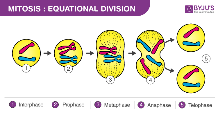
Mitosis strictly refers only to the division of the genetic material in the nucleus (Karyokinesis). The ultimate goal is flawless genetic fidelity: producing two genetically identical diploid (2n) daughter cells from a single diploid parent cell.


Detailed Cellular Mechanics of Mitosis
The Detailed Sequence
-
Prophase
Condensation: Chromatin coils into visible chromosomes (sister chromatids joined by Cohesins). Nucleolus disappears. The mitotic spindle begins to form. -
Prometaphase
Envelope Breakdown: Nuclear envelope fragments. Microtubules attach to the Kinetochores at the centromeres. -
Metaphase
Alignment: Chromosomes align on the Metaphase Plate. The cell halts at the Spindle Assembly Checkpoint until all kinetochores are under tension. -
Anaphase
Separation: APC/C unleashes Separase, which cleaves cohesins. Sister chromatids are instantly pulled to opposite poles. -
Telophase & Cytokinesis
Reconstruction: Nuclear envelopes rebuild. Chromosomes decondense. The cytoplasm divides via an actin-myosin cleavage furrow (animals) or a cell plate (plants).
🧬 3. Meiosis: Sexual Reproduction & Genetic Variation

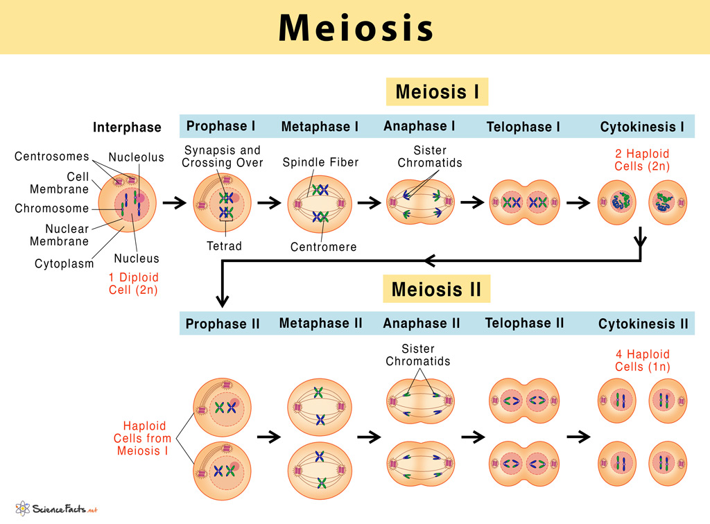
Meiosis: Homologous chromosomes separate in meiosis I and sister chromatids separate in meiosis II, producing four genetically varied haploid cells from one diploid precursor.

Crossing over: Homologues synapse during prophase I and nonsister chromatids exchange corresponding DNA at chiasmata, generating recombinant chromosomes.
Meiosis requires two consecutive cell divisions (Meiosis I and II) following a single round of DNA replication. It reduces the chromosome count from diploid (2n) to haploid (n) to ensure fertilization restores the normal diploid state.


Meiosis I: Crossing Over & Independent Assortment
- Prophase I: Homologs pair up (Synapsis) forming tetrads. Crossing Over occurs at chiasmata, physically exchanging DNA to create recombinant chromosomes.
- Metaphase I: Tetrads align randomly (Independent Assortment), creating $2^{23}$ possible combinations in humans.
- Anaphase I: Homologs separate. Shugoshin protects centromeric cohesins, so sister chromatids stay together. Halves chromosome number.
- Mechanically identical to Mitosis, but starting with haploid (n) cells.
- Anaphase II: Centromeric cohesins are cleaved. Sister chromatids finally separate.
- Result: Four genetically unique haploid gametes.
⚖️ 4. Mitosis vs. Meiosis Summary

| Feature | Mitosis | Meiosis |
|---|---|---|
| Purpose | Growth, tissue repair, asexual reproduction. | Gamete production for sexual reproduction. |
| Divisions | One | Two |
| Crossing Over? | No. | Yes (Prophase I). |
| Final Outcome | 2 identical Diploid (2n) cells | 4 distinct Haploid (n) cells |
🚨 5. Clinical Pathology & Cancer

Apoptosis: Caspase-mediated programmed death causes cell shrinkage, chromatin condensation, membrane blebbing, and apoptotic bodies that are cleared without major inflammation.

Apoptosis versus necrosis: Apoptosis is controlled and membrane integrity is largely retained; necrosis involves swelling, rupture, leakage, and an inflammatory response.

Multistep cancer development: Accumulated activation of proto-oncogenes and loss of tumour-suppressor or DNA-repair genes progressively remove controls on proliferation and survival.

Benign versus malignant: Benign tumours remain localised; malignant cells invade surrounding tissue and may enter blood or lymph to establish metastases.


Nondisjunction is the failure of chromosomes to separate properly.
• Meiosis I Error: Homologs fail to separate → 100% abnormal gametes.
• Meiosis II Error: Sister chromatids fail to separate → 50% normal, 50% abnormal gametes.
- Trisomy 21 (Down Syndrome): 47, XX,+21. Most common viable trisomy.
- Trisomy 18 (Edwards) / 13 (Patau): Severe developmental issues, usually fatal.
- Klinefelter (47, XXY): Male, sterile. 1 Barr body.
- Turner (45, X0): Female. Only viable human monosomy. 0 Barr bodies.
Structural Abnormalities & Cancer
Translocation: Movement of a segment to a non-homologous chromosome. E.g., The Philadelphia Chromosome ($t(9;22)$) fuses BCR-ABL, causing Chronic Myelogenous Leukemia (CML).
Cancer Genetics: Cancer bypasses checkpoints. Oncogenes (like mutated Ras) are dominant "gas pedals". Tumor Suppressors (like p53 or Rb) are recessive "brakes" requiring two hits to fail.
🌱 6. Reproduction and Heredity

Stem-cell potency: Totipotent cells can form embryonic and extraembryonic tissues; pluripotent cells form all body cell types; multipotent and unipotent cells have progressively narrower fates.

Stem-cell niche: Local signals maintain self-renewal. Asymmetric division can preserve one stem cell while producing one committed progenitor for tissue renewal.
Reproduction ensures the survival of a species. It manifests in two fundamentally different strategies across the tree of life.
Asexual Reproduction
A single parent produces genetically identical clones via mitosis or binary fission. Energy efficient and rapid, but lacks genetic variation, making populations vulnerable to environmental shifts.
- Binary Fission: Bacteria/Archaea splitting.
- Budding: Yeast, Hydra.
- Fragmentation: Starfish, Planaria.
- Parthenogenesis: Unfertilized egg develops into an adult (Bees, some lizards).
Sexual Reproduction
Two parents contribute gametes (produced via meiosis) that fuse during fertilization to form a unique diploid zygote. Energy intensive, but creates massive genetic variation crucial for evolution and adaptation.

Sexual Life Cycles
All sexual life cycles alternate between meiosis (halving chromosome count) and fertilization (doubling it). The timing varies by kingdom:
Diagram 6.1: The Three Sexual Life Cycles
One cycle connects every term in this chapter
Use the color code throughout the section: germline → male gamete / female gamete → fertilization → development.
$2n$ → meiosis
$n$
$n$
$n+n\rightarrow2n$
6.1 Germ Cells, Germline, and Gametes
A germ cell is a cell belonging to the lineage that can ultimately transmit genetic information to the next generation. In animals, the germline is separated conceptually from the somatic cell lineage, which forms the tissues of the body. Mature germ cells are the gametes: spermatozoa in males and the ovulated secondary oocyte/ovum in females. Importantly, not every germ cell is already a gamete; spermatogonia, oogonia, primary spermatocytes, and primary oocytes are all germ-line cells at earlier developmental stages.
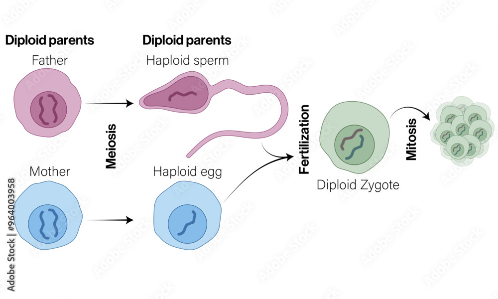

Diagram 6.2: The Human Germline Cycle
6.2 Chromosome Number ($n$) vs. DNA Content ($C$)
A frequent exam trap is confusing chromosome number with DNA amount. The symbol $n$ describes how many homologous chromosome sets are present, whereas $C$ describes the amount of DNA relative to one haploid genome. DNA replication doubles $C$ without changing $n$ because sister chromatids remain joined at one centromere and are still counted as one chromosome.
| Cell / stage | Ploidy | DNA content | Key interpretation |
|---|---|---|---|
| Spermatogonium / oogonium in G$_1$ | $2n$ | $2C$ | Diploid, chromosomes not yet replicated. |
| Primary spermatocyte / primary oocyte after S phase | $2n$ | $4C$ | Still diploid, but every chromosome contains two sister chromatids. |
| Secondary spermatocyte / secondary oocyte | $n$ | $2C$ | Homologues separated in meiosis I; sister chromatids remain joined. |
| Spermatid / ovum after meiosis II | $n$ | $1C$ | Sister chromatids have separated. |
| Zygote after fusion of pronuclei | $2n$ | $2C$ | Maternal and paternal haploid genomes are combined before the first S phase. |
6.3 Spermatogenesis: Production of Male Gametes
Spermatogenesis occurs in the seminiferous tubules of the testes and begins functionally at puberty. One diploid primary spermatocyte ultimately produces four functional haploid spermatozoa because cytokinesis is approximately equal during the meiotic divisions.
Diagram 6.3: Spermatogenesis — Ploidy and Differentiation
- Spermatogonia: diploid stem/progenitor cells near the basal compartment of seminiferous tubules. Mitotic divisions maintain the germ-line pool and generate cells committed to meiosis.
- Primary spermatocyte: enters meiosis I after DNA replication. Homologous chromosomes synapse, cross over, and then separate.
- Secondary spermatocytes: haploid with duplicated chromosomes. They rapidly enter meiosis II.
- Spermatids: haploid products of meiosis II. They are not yet mature motile sperm.
- Spermiogenesis: differentiation of spermatids into spermatozoa without another cell division. The nucleus condenses, an acrosome forms, mitochondria concentrate in the midpiece, and a flagellum develops.
- Spermiation: release of mature spermatozoa from Sertoli cells into the lumen of seminiferous tubules.
Sertoli Cells
Support and nourish developing germ cells, form the blood-testis barrier, respond to FSH and testosterone, phagocytose residual cytoplasm, and secrete inhibin B to reduce FSH secretion.
Leydig Cells
Located in the interstitial tissue between seminiferous tubules. They respond primarily to LH and synthesize testosterone, which supports spermatogenesis and male secondary sexual characteristics.
6.4 Structure and Functional Adaptations of Sperm
6.5 Oogenesis: Production of Female Gametes
Oogenesis is highly asymmetric. Cytokinesis preserves almost all cytoplasm in one cell, producing a single large functional gamete and small polar bodies. In humans, the meiotic program begins before birth, pauses for years, resumes cyclically after puberty, and is completed only if fertilization occurs.
Diagram 6.4: Oogenesis — Arrest Points Are High-Yield
- Oogonia: proliferate by mitosis during fetal development and give rise to primary oocytes.
- Primary oocyte: begins meiosis I before birth and arrests in Prophase I (classically the diplotene/dictyate stage).
- Completion of meiosis I: around ovulation, a selected primary oocyte divides asymmetrically into a large secondary oocyte and a small first polar body.
- Secondary oocyte: enters meiosis II and arrests at Metaphase II. This is the cell typically ovulated in humans.
- Fertilization: sperm entry activates the oocyte and normally triggers completion of meiosis II, producing the ovum and another polar body.
6.6 Folliculogenesis and the Ovarian Cycle
| Stage / phase | Major event | Key cells / hormones | High-yield point |
|---|---|---|---|
| Primordial follicle | Primary oocyte surrounded by flattened follicular cells. | Quiescent ovarian reserve | Primary oocyte remains arrested in Prophase I. |
| Growing primary/secondary follicle | Granulosa cells proliferate; zona pellucida develops; theca differentiates. | FSH supports granulosa cells; LH stimulates theca androgen production. | Granulosa aromatase converts androgens to estrogens. |
| Antral / dominant follicle | Fluid-filled antrum enlarges and one follicle becomes dominant. | Rising estradiol | Sustained high estradiol switches to positive feedback and promotes the LH surge. |
| Ovulation | Secondary oocyte is released from the ovary. | LH surge | Ovulated cell is generally arrested in Metaphase II. |
| Corpus luteum | Post-ovulatory follicle becomes a temporary endocrine gland. | Progesterone, estrogen, inhibin | Progesterone supports a secretory endometrium. |
| If pregnancy occurs | Early embryo maintains corpus luteum. | hCG from trophoblast | hCG acts in an LH-like manner to support progesterone secretion until placental steroid production is sufficient. |
6.7 Fertilization: From Gamete Recognition to Zygote
Human fertilization normally occurs in the ampulla of the uterine tube. It is not merely the physical collision of two gametes; it is a coordinated sequence of activation, recognition, membrane fusion, prevention of polyspermy, and union of the two haploid genomes.
Diagram 6.5: Fertilization Sequence
- Capacitation: sperm membranes are biochemically modified in the female tract, increasing motility patterns and preparing the sperm for the acrosome reaction.
- Corona radiata penetration: sperm moves through the follicular cells surrounding the oocyte.
- Zona pellucida interaction and acrosome reaction: sperm-oocyte recognition triggers release/exposure of acrosomal components needed for further penetration.
- Plasma membrane fusion: sperm and oocyte membranes fuse. The oocyte is activated.
- Cortical reaction / zona reaction: cortical granules release their contents and modify the zona pellucida, strongly reducing the ability of additional sperm to enter. This is a major block to polyspermy.
- Completion of meiosis II: the secondary oocyte finishes meiosis II and extrudes the second polar body.
- Pronuclear formation and syngamy: paternal and maternal genomes form pronuclei and then combine functionally to establish the diploid zygotic genome.
6.8 Oocyte Coverings and Fertilization Vocabulary
6.9 From Zygote to Implantation: Essential Early-Development Terms
After fertilization, the zygote undergoes repeated mitotic divisions called cleavage. Cell number increases rapidly while total embryonic size changes relatively little at first.
| Term | Meaning | Why it matters |
|---|---|---|
| Cleavage | Rapid mitotic divisions of the early embryo. | Produces progressively smaller cells called blastomeres without an equivalent increase in total embryo size. |
| Blastomere | One cell produced by cleavage of the zygote. | Early blastomeres contribute to the embryo and supporting structures. |
| Morula | Compact solid ball of blastomeres. | Precedes formation of a fluid-filled blastocyst. |
| Blastocyst | Early mammalian embryo containing a cavity, trophoblast, and inner cell mass. | Stage that attaches to and begins implantation in the uterine endometrium. |
| Trophoblast | Outer cell layer of the blastocyst. | Contributes importantly to implantation and fetal components of the placenta; early trophoblastic tissue produces hCG. |
| Inner cell mass / embryoblast | Cluster of cells inside the blastocyst. | Gives rise to the embryo proper. |
| Implantation | Attachment and invasion of the blastocyst into the uterine endometrium. | Establishes intimate maternal-embryonic contact needed for continued development. |
| Gastrulation | Major reorganization that establishes the three primary germ layers. | Creates the basic body plan and the precursors of all major tissues. |
| Ectoderm | Outer germ layer. | Major derivatives include epidermis and nervous system. |
| Mesoderm | Middle germ layer. | Major derivatives include muscle, bone, connective tissue, blood, heart, kidneys, and gonads. |
| Endoderm | Inner germ layer. | Major derivatives include epithelial linings of much of the digestive and respiratory systems and associated organs. |
6.10 Endocrine Control of Human Reproduction
Diagram 6.6: Hypothalamic–Pituitary–Gonadal Axis
- GnRH: released from the hypothalamus in pulses and stimulates the anterior pituitary.
- FSH: supports Sertoli-cell function and spermatogenesis in males; promotes follicular/granulosa-cell development in females.
- LH: stimulates Leydig cells to produce testosterone in males; in females it supports theca-cell steroidogenesis, triggers ovulation during the LH surge, and supports corpus luteum formation.
- Inhibin: produced by Sertoli or granulosa cells and selectively suppresses FSH secretion.
- Estrogens: promote growth and function of female reproductive tissues and participate in feedback regulation of the hypothalamic-pituitary axis.
- Progesterone: especially important after ovulation; converts/maintains the endometrium in a secretory state favorable for implantation.
- Testosterone: supports spermatogenesis and male reproductive differentiation and secondary sexual characteristics.
6.11 High-Yield Comparison: Spermatogenesis vs. Oogenesis
| Feature | Spermatogenesis | Oogenesis |
|---|---|---|
| Primary site | Seminiferous tubules of testes | Ovarian follicles |
| Functional onset in humans | Mainly from puberty onward | Meiosis begins before birth; cyclic maturation resumes after puberty |
| Major arrest points | No long physiologic meiotic arrest comparable with oocytes | Primary oocyte: Prophase I; secondary oocyte: Metaphase II |
| Cytokinesis | Approximately equal | Highly unequal |
| Functional products from one primary meiotic cell | Four spermatozoa | One large functional ovum plus polar bodies |
| Gamete size/motility | Small and motile | Large and non-motile; contains abundant cytoplasm and organelles |
| Final meiotic completion | Completed before mature sperm function | Meiosis II completes only after fertilization in the normal human cycle |
6.12 Extended Reproduction & Development Glossary
- A cell after DNA replication is not automatically tetraploid. A human primary spermatocyte is still $2n$ but has $4C$ DNA.
- Meiosis I separates homologous chromosomes; meiosis II separates sister chromatids.
- The human cell released at ovulation is normally a secondary oocyte arrested in Metaphase II, not a fully completed ovum.
- One primary spermatocyte can yield four functional sperm; one primary oocyte yields only one large functional female gamete because cytokinesis is asymmetric.
- Fertilization restores the chromosome-set number from $n+n$ to $2n$; it does not create a tetraploid cell.
| Feature | Spermatogenesis | Oogenesis |
|---|---|---|
| Starts | At puberty from spermatogonial stem cells | Primary oocytes are formed before birth and arrest in Prophase I |
| Cytokinesis | Approximately equal | Highly unequal |
| Products per primary cell | Four functional sperm | One large functional ovum + polar bodies |
| Meiotic arrest | No prolonged physiological arrest comparable to oogenesis | Prophase I, then Metaphase II |
| Completion | Continuous after puberty | Meiosis II completes only after fertilization |
| Cell specialization | Spermiogenesis creates acrosome, compact nucleus, flagellum | Large cytoplasm stores organelles, RNAs and developmental resources |
🧬 7. Molecular Genetics: DNA Structure & Replication

Semiconservative replication: The parental strands separate and each templates a complementary daughter strand, so every product contains one old strand and one newly synthesised strand.
DNA Structure (Review)
DNA is a double-stranded, antiparallel right-handed helix. The backbone is formed by alternating deoxyribose sugars and phosphate groups linked by phosphodiester bonds ($5' \rightarrow 3'$). The rungs are nitrogenous bases linked by hydrogen bonds (A=T with 2 bonds; G$\equiv$C with 3 bonds).

DNA Replication
Replication is semi-conservative (each new molecule has one old strand and one new strand) and occurs during the S phase.
Diagram 7.1: The DNA Replication Fork
The Enzymatic Arsenal
- Helicase: Unzips the double helix by breaking H-bonds.
- Single-Strand Binding Proteins (SSBPs): Prevent strands from re-annealing.
- Topoisomerase (Gyrase): Relieves supercoiling tension ahead of the fork.
- Primase: Lays down a short RNA primer (DNA Pol requires an existing 3'-OH).
- DNA Polymerase III: Synthesizes the new DNA strand only in the 5' → 3' direction.
- DNA Polymerase I: Replaces RNA primers with DNA.
- DNA Ligase: Seals nicks in the phosphodiester backbone between Okazaki fragments on the lagging strand.
🧬 8. The Central Dogma: Transcription & Translation
The Central Dogma states that information flows from DNA → RNA → Protein. The Genetic Code translates 3-nucleotide sequences (Codons) into amino acids. It is universal, unambiguous, and degenerate (redundant).
Start Codon: AUG (Methionine) | Stop Codons: UAA, UAG, UGA

1. Transcription (DNA to mRNA)
Occurs in the nucleus (eukaryotes) or nucleoid (prokaryotes). RNA Polymerase binds to the Promoter (e.g., TATA box), separates DNA, and synthesizes a complementary mRNA strand $5' \rightarrow 3'$ off the template strand.
Before leaving the nucleus, pre-mRNA gets a 5' GTP Cap (protection/ribosome attachment), a 3' Poly-A Tail (protection/export), and undergoes Splicing (Spliceosomes remove non-coding introns and join coding exons).
2. Translation (mRNA to Protein)
Occurs at the Ribosome in the cytoplasm. Transfer RNA (tRNA) brings specific amino acids based on mRNA codons.
Diagram 8.1: Translation at the Ribosome
- A Site (Aminoacyl): Binds incoming charged tRNA matching the codon.
- P Site (Peptidyl): Holds the tRNA carrying the growing polypeptide chain. A peptide bond forms between this chain and the amino acid in the A site.
- E Site (Exit): Uncharged tRNA exits the ribosome.
🧬 9. Regulation of Gene Expression

Lac operon: Without lactose the repressor occupies the operator. Allolactose inactivates the repressor; when glucose is also low, cAMP–CAP strongly promotes transcription.

Eukaryotic regulation: Activators bind enhancers and DNA looping brings them near promoter-bound general transcription factors and RNA polymerase II. Chromatin accessibility adds another regulatory layer.
Prokaryotic Regulation: The Operon Model
Bacteria regulate genes functionally related into single units called Operons. They are transcribed into a single polycistronic mRNA.
- Promoter: Where RNA Polymerase binds.
- Operator: The "on/off switch" within the promoter.
- Repressor: A protein that binds the operator, physically blocking RNA Polymerase.
- Inducible Operon (e.g., lac operon): Usually OFF. An inducer (allolactose) binds the repressor, removing it, turning the operon ON.
- Repressible Operon (e.g., trp operon): Usually ON. A corepressor (tryptophan) binds the repressor, activating it to bind the operator, turning it OFF.
Eukaryotic Regulation: Complex Control
Eukaryotic control is multi-tiered and much more complex than prokaryotes.
- Histone Acetylation: Loosens chromatin (Euchromatin) → Increases transcription.
- DNA Methylation: Tightens chromatin (Heterochromatin) → Decreases/silences transcription.
Requires Transcription Factors. Specific activators bind to distal Enhancer DNA sequences, looping the DNA to help RNA Polymerase initiate at the promoter.
| System | Default state | Signal | Outcome |
|---|---|---|---|
| lac operon | Repressor blocks operator | Allolactose + low glucose favor expression | Genes for lactose utilization are transcribed |
| trp operon | Transcription possible when tryptophan is scarce | High tryptophan acts as corepressor | Repressor binds operator and reduces transcription |
| Eukaryotic enhancer | Depends on chromatin state | Specific transcription factors | Promoter recruitment and transcription increase |
| RNA interference | Target mRNA present | miRNA/siRNA-guided RISC | Translation repression or mRNA cleavage/degradation |
🎨 10. High-Yield Visual Glossary & Integrative Review
This final review does not replace the detailed sections above. It compresses the most testable vocabulary into color-coded retrieval cues so you can revise by recognition first, then return to the full explanations when a term feels weak.
Mastery Practice Quiz
Test your comprehensive understanding of the cell cycle, mitosis, meiosis, molecular genetics, and clinical pathology. These 25 high-yield questions are styled after the rigorous, integrated logic required for the IMAT examination and now include germ cells, spermatogenesis, oogenesis, fertilization, and early development.