Which of the following rows are correct about these naturally occurring biological molecules?
Row
biological molecule
presence of carboxylic acid groups
presence of 2 or more hydroxyl groups
1
ALL amino acids
yes
no
2
glucose
no
yes
3
glycerol
no
yes (Corrected from 'no' based on glycerol's 3 OH groups)
4
ALL fatty acids
yes
no
IMAT Challenge
Question 2 Challenge
2012 - Q58
Which of the following is/are true about hydrogen bonds between water molecules? 1 They are weak bonds. 2 They are strong bonds. 3 They are temporary bonds. 4 They require hydrolysis to break.
IMAT Challenge
Question 1 Challenge
2024 - Q11
What is a hydrogen bond?
Biological macromolecules are massive, highly complex organic molecules that construct the physical architecture and the functional, catalytic machinery of all living organisms. The vast majority of these macromolecules are polymers—long, repeating chains assembled from smaller, simpler molecular building blocks known as monomers. The process of linking these monomers together is one of the most fundamental reactions in all of biochemistry.
The fundamental concept of biochemistry: small, simple organic monomers (like monosaccharides or amino acids) are covalently linked together in long chains to form massive, functionally diverse polymers (like starch or proteins).
The Universal Chemistry of Polymerization
Regardless of whether the cell is synthesizing a carbohydrate, a protein, or a nucleic acid, the chemical mechanisms underlying the assembly and disassembly of these polymers revolve entirely around the molecule of Water ($H_2O$).
1. Condensation (Dehydration Synthesis)
This is an anabolic process. Two separate monomers are covalently joined together to form a larger polymer. In the process, a hydroxyl group ($-OH$) is removed from one monomer, and a hydrogen atom ($-H$) is removed from the other. These immediately combine to form and release a molecule of water.
Because building complex, highly ordered bonds from free-floating monomers decreases the entropy of the system, this process is strictly endergonic and always requires an input of energy (usually supplied by the hydrolysis of ATP).
This is a catabolic process. It is the exact chemical reverse of condensation. To break the covalent bond holding a polymer together, a molecule of water is forcefully inserted (lysed) across the bond, restoring the $-OH$ and $-H$ to the respective individual monomers, thereby separating them.
This process increases entropy and is therefore highly exergonic, releasing the energy stored in the chemical bond. It is the fundamental biochemical mechanism behind all food digestion in the human gastrointestinal tract.
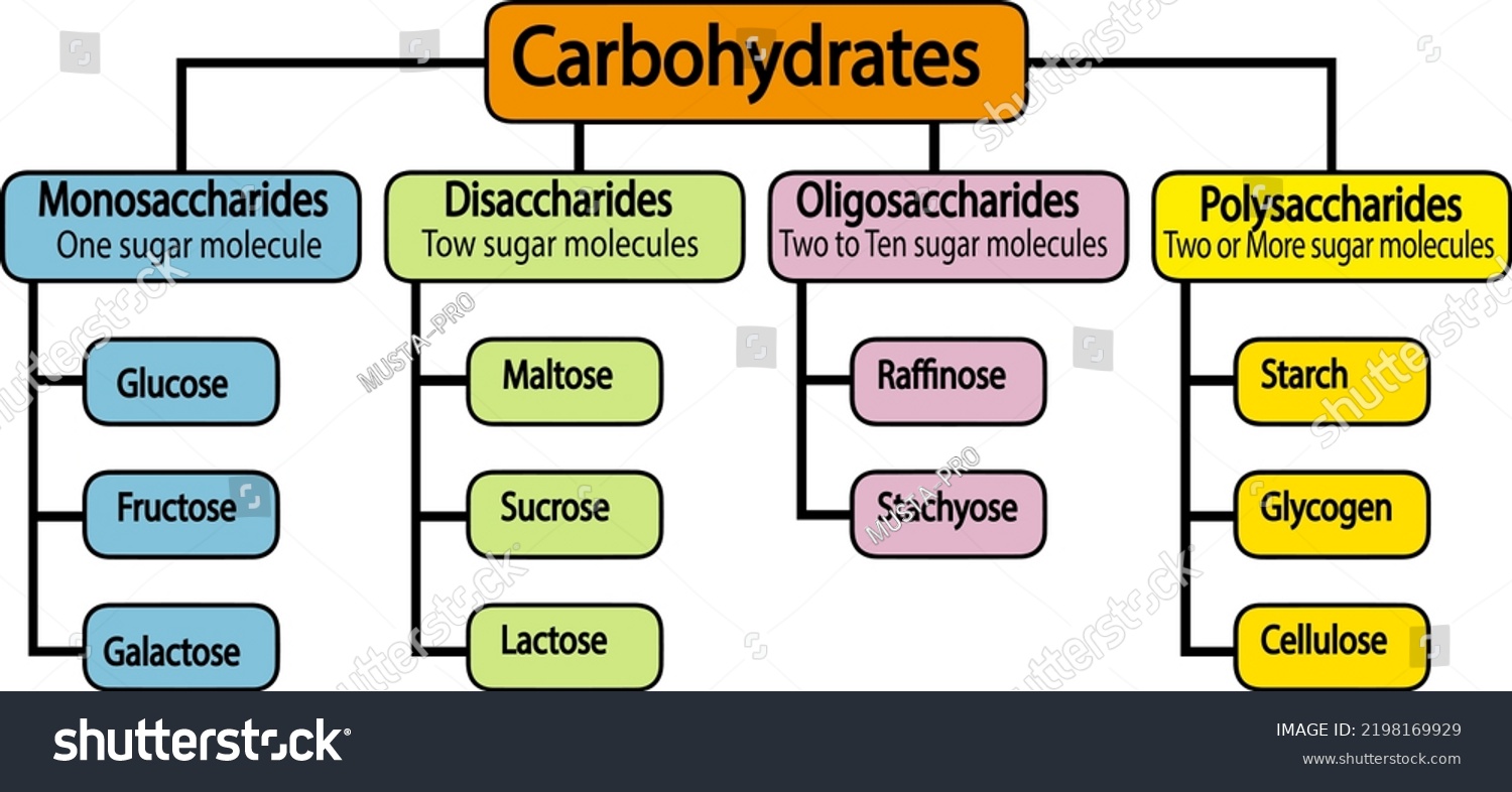
Carbohydrate classification: a visual overview of mono-, di-, oligo- and polysaccharide categories. Use it as a map before studying the individual structures below.
IMAT Challenge
Question 3 Challenge
2024 - Q13
What kind of monosaccharide is glucose?
Carbohydrates are polyhydroxy aldehydes or ketones. Historically named because their chemical formulas make them appear as "hydrates of carbon", they are composed exclusively of Carbon, Hydrogen, and Oxygen atoms, almost universally obeying the strict stoichiometric ratio of $1:2:1$. This is represented by the empirical formula $(CH_2O)_n$. They are the most abundant class of biomolecules on Earth, acting as the primary fuel source for cellular respiration and as robust structural components.
Visual examples of the different classes of carbohydrates, showing their immense structural diversity ranging from simple, water-soluble monosaccharides up to massive, insoluble structural polysaccharides.
Monosaccharides (Simple Sugars)
Monosaccharides are the simplest, indivisible monomeric units of carbohydrates. Due to the presence of multiple highly polar, electronegative hydroxyl ($-OH$) functional groups along their carbon chain, they form extensive hydrogen bonds with water. This makes them extremely soluble in aqueous solutions, forming sweet-tasting, crystalline solids at room temperature. They are classified biochemically by two primary metrics: the total number of carbon atoms they possess, and whether their defining carbonyl group ($C=O$) is an aldehyde (located at the end of the chain) or a ketone (located internally).
Class Name
Carbons
Chemical Formula
Key Biological Examples & Functions
Triose
3
$C_3H_6O_3$
Glyceraldehyde (G3P): An absolutely crucial 3-carbon intermediate metabolite found universally in both cellular respiration (specifically within glycolysis) and photosynthesis (as the direct product of the Calvin Cycle).
Pentose
5
$C_5H_{10}O_5$
Ribose: The structural 5-carbon backbone of RNA, ATP, and essential electron carriers like $NAD^+$. It possesses a critical, reactive hydroxyl group ($-OH$) at the Carbon-2 position.
Deoxyribose ($C_5H_{10}O_4$): The structural backbone of DNA. As the name heavily implies ("De-oxy"), it lacks the oxygen atom at the Carbon-2 position, containing only a Hydrogen atom there instead. This lack of a highly reactive oxygen makes the DNA molecule vastly more chemically stable and resistant to hydrolysis than RNA.
Hexose
6
$C_6H_{12}O_6$
Glucose (Aldose): The primary blood sugar in humans and the universal fuel for cellular aerobic respiration.
Fructose (Ketose): Fruit sugar. The sweetest naturally occurring sugar, processed primarily in the liver.
Galactose (Aldose): A structural component of mammalian milk sugar (lactose). It cannot be directly utilized for energy by the brain; it must first be enzymatically converted to glucose in the liver.
High Yield IMAT Concept - Stereochemistry
Deep Dive: Isomerism & Molecular Chirality
Because biological enzymes are staggeringly specific 3D protein structures, the exact spatial orientation of atoms in a substrate is just as important as its raw chemical formula. Monosaccharides exhibit multiple, overlapping layers of complex isomerism.
1. Structural Isomers (Aldoses vs. Ketoses)Glucose, Galactose, and Fructose all share the exact same empirical chemical formula: $C_6H_{12}O_6$. However, they differ radically in how their atoms are bonded. Glucose and Galactose possess an aldehyde group at Carbon-1 (making them Aldohexoses). Fructose, however, possesses a ketone group at Carbon-2 (making it a Ketohexose). This fundamental difference dictates ring formation: Fructose typically forms a 5-membered furanose ring, while Glucose normally forms a 6-membered pyranose ring in solution.
2. Optical Isomers (Enantiomers & Epimers)Any carbon atom covalently bonded to four completely different chemical groups is termed a chiral center. This chirality creates non-superimposable mirror images of the molecule (enantiomers, D vs L). By universal biochemical convention, living biological systems have evolved to almost exclusively synthesize and metabolize D-Isomer carbohydrates (e.g., D-Glucose).
Epimers are a highly specific type of diastereomer that differ in spatial configuration at exactly one specific chiral carbon. For instance, D-Glucose and D-Galactose are C-4 epimers. They are structurally identical everywhere except that the $-OH$ group on Carbon-4 points in the opposite spatial direction. This tiny geometric flip makes Galactose completely unrecognizable to the enzyme Hexokinase.
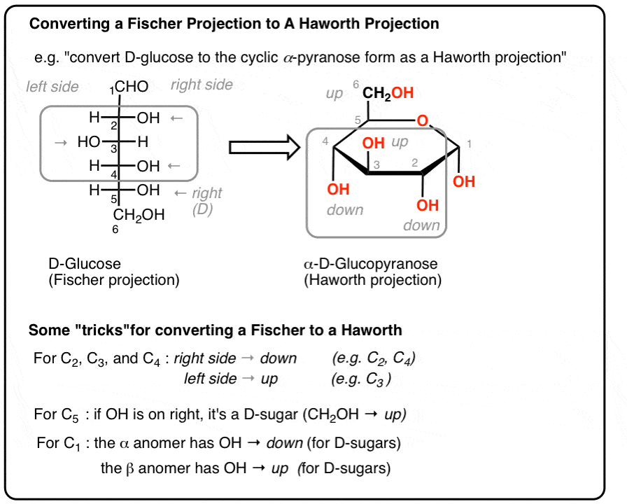
3. Anomers ($\alpha$ vs $\beta$ configurations) & MutarotationWhen a linear, straight-chain monosaccharide drops into an aqueous solution, it violently attacks itself (the hydroxyl group on C-5 attacks the carbonyl carbon at C-1). This intramolecular nucleophilic attack forms a cyclic ring (a hemiacetal). Crucially, this ring closure transforms Carbon-1 from an achiral carbon into a brand new chiral center, which is formally called the Anomeric Carbon. The new $-OH$ group on this anomeric carbon can lock into one of two distinct positions relative to the bulky $CH_2OH$ group at C-6:
$\alpha$ (Alpha) configuration:
The $-OH$ group points DOWN (Trans to C-6).
$\beta$ (Beta) configuration:
The $-OH$ group points UP (Cis to C-6).
Thermodynamics of Mutarotation: In an aqueous solution, the ring rapidly opens and closes, constantly flipping back and forth between the $\alpha$ and $\beta$ forms in a dynamic process known as Mutarotation until thermodynamic equilibrium is reached (roughly 36% $\alpha$, 64% $\beta$, because the equatorial $\beta$ position experiences less physical steric hindrance).
Detailed animation showing the nucleophilic attack responsible for the cyclization of D-glucose from its linear (Fischer projection) form into its highly stable, cyclic ring (Haworth projection) form. Note the creation of the brand new anomeric carbon at C-1 upon ring closure.
Structural Visualization: The Anomers of Glucose
🔢 Carbon-number vocabulary
Triose = 3 C, pentose = 5 C, hexose = 6 C. The carbonyl position then distinguishes an aldose from a ketose.
🪞 D/L configuration
D and L describe the configuration of the highest-numbered chiral carbon relative to glyceraldehyde. They do not mean dextrorotatory and levorotatory.
🎯 Epimer
Epimers differ at exactly one chiral carbon. Glucose–galactose differ at C4; glucose–mannose differ at C2.
🔁 Anomer
Ring closure creates the anomeric carbon. $\alpha$ and $\beta$ forms differ only at this carbon and interconvert in solution by mutarotation.
🧪 Reducing sugar
A free anomeric carbon can open to a reactive carbonyl form and reduce Benedict/Fehling reagent. Most monosaccharides are reducing sugars.
🧬 Ribose vs deoxyribose
Ribose has an $-OH$ at C2; deoxyribose has H instead. That single oxygen difference contributes to DNA's greater chemical stability.
🍩 2. Disaccharides: Double Sugars
Disaccharides are constructed by the enzymatic, endergonic condensation (dehydration synthesis) of two specific monosaccharides. The resulting strong covalent bond holding the two sugars together is called a Glycosidic Bond. Like all condensation reactions, the formation of this bond results in the loss of one molecule of water.
General Stoichiometric Equation
$C_6H_{12}O_6 + C_6H_{12}O_6 \longrightarrow C_{12}H_{22}O_{11} + H_2O$
Mechanism: Glycosidic Bond Formation
The most common linkage in biology occurs between the highly reactive anomeric carbon (Carbon-1) of the first sugar molecule and the relatively stable Carbon-4 of the second sugar molecule. This specific geometry creates a linear 1 → 4 Glycosidic Linkage.
Biochemical Mechanism: Condensation Synthesis of Maltose
Produced in massive quantities during the digestion of starch by the enzyme Amylase (found in saliva and pancreatic juice). It retains a free anomeric carbon, making it a highly reactive Reducing Sugar.
The standard table sugar extracted from sugar cane. It is the primary transport sugar in the phloem sap of plants precisely because the unique bond locks both anomeric carbons, rendering it exceptionally stable and unreactive (Non-Reducing Sugar).
The primary sugar found in mammalian milk, providing dense energy for nursing infants. Its beta bond makes it difficult to digest, requiring a highly specific enzyme to break. It is a Reducing Sugar.
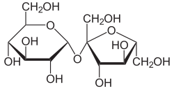
The chemical structure of Sucrose (Table Sugar). Observe the bizarre $\alpha(1 \rightarrow 2)\beta$ glycosidic linkage permanently bridging the glucose pyranose ring with the fructose furanose ring, locking both reactive anomeric carbons.
Deep Concept: Why is Sucrose "Non-Reducing"?
A "reducing sugar" is chemically defined as any carbohydrate capable of acting as a reducing agent. To do this, the sugar must possess a "free" anomeric carbon capable of momentarily opening its ring structure in aqueous solution to expose a highly reactive aldehyde or ketone functional group.
In maltose and lactose, the glycosidic bond only utilizes Carbon-1 of the first sugar. The anomeric carbon on the second sugar remains untouched and "free" to open, react, and reduce other chemicals (like the copper in Benedict's reagent).
The Sucrose Exception: In Sucrose, the condensation reaction occurs directly between Carbon-1 of glucose AND Carbon-2 of fructose. Because both of these are the anomeric carbons of their respective rings, both rings are permanently locked shut by the bond. Neither can open to form an aldehyde/ketone. Therefore, sucrose is chemically inert and is termed a Non-Reducing Sugar.
Clinical Correlates - Gastroenterology
Lactose Intolerance
Mammals synthesize the enzyme Lactase as infants specifically to cleave the stubborn $\beta(1 \rightarrow 4)$ glycosidic bond of lactose. In most humans worldwide (except populations with a specific evolutionary mutation), lactase gene expression plummets after weaning. If a lactase-deficient adult consumes dairy, the intact, bulky lactose travels undigested into the large intestine.
Pathology: The heavy lactose molecules wildly increase the osmolarity of the colon, drawing massive amounts of water out of the blood and into the gut via osmosis, causing severe, explosive diarrhea. Simultaneously, opportunistic anaerobic gut bacteria rapidly ferment the lactose, producing excessive $CO_2$, methane, and hydrogen gas, leading to painful bloating and flatulence.
Galactosemia
A severe, potentially fatal autosomal recessive genetic metabolic disorder. Infants born with this disease lack the specific enzymes (most commonly GALT) required to convert the galactose (derived from the breakdown of milk lactose) into usable glucose.
Pathology: Toxic levels of galactose and its heavily reduced, highly osmotic metabolite (galactitol) rapidly accumulate in the blood and tissues. This buildup causes catastrophic liver damage (hepatomegaly and cirrhosis), profound mental retardation, and the rapid, irreversible formation of bilateral cataracts in the eyes of newborns within days of drinking milk.
🍞 Maltose
Glucose + glucose, usually $\alpha(1\rightarrow4)$. Produced during starch digestion; hydrolysed by maltase; reducing because one anomeric carbon remains free.
🥛 Lactose
Galactose + glucose, $\beta(1\rightarrow4)$. Hydrolysed by lactase. Lactase deficiency causes osmotic diarrhea and bacterial fermentation.
🍬 Sucrose
Glucose + fructose, $\alpha1\leftrightarrow\beta2$. Both anomeric carbons participate in the bond, so sucrose is non-reducing until hydrolysed by acid or sucrase.
🌳 3. Polysaccharides: Complex Polymers
IMAT Challenge
Question 19 Challenge
2012 - Q41
If a glucose molecule became incorporated as a non-terminal component of starch, which two regions, labelled i to v, would be involved in forming glycosidic bonds?
IMAT Challenge
Question 17 Challenge
2013 - Q33
Which one of the following does not contain amino acids?
Polysaccharides are massive, towering macromolecules composed of hundreds to many thousands of monosaccharides linked by glycosidic bonds. Unlike simple sugars, they are entirely insoluble in water (meaning they are osmotically neutral and do not draw water into the cell, which is absolutely crucial for safe intracellular storage) and they are completely tasteless. Their macroscopic biological function—whether they act as a cellular battery (energy storage) or as physical building materials (structural support)—is determined entirely by the specific isomer of glucose they are built from ($\alpha$ vs $\beta$) and the specific geometry of their glycosidic branching.
A. Energy Storage Polymers (Built exclusively from $\alpha$-Glucose)
1. Starch (Plants)
Plants store their excess glucose manufactured during photosynthesis as dense starch granules within specialized plastids called amyloplasts. Starch is a heterogeneous mixture of two distinct polymers:
Amylose (approx. 20%): An unbranched, perfectly linear chain. Consists exclusively of $\alpha(1 \rightarrow 4)$ bonds. Due to bond angles, it naturally coils into a tight helical spring, making it incredibly compact but slow to digest (enzymes can only attack the two ends).
Amylopectin (approx. 80%): A highly complex, branched polymer. Contains the $\alpha(1 \rightarrow 4)$ backbone plus $\alpha(1 \rightarrow 6)$ branch points. Faster hydrolysis due to numerous terminal branches.
2. Glycogen (Animals)
Stored in massive, dense granules primarily within the hepatocytes of the Liver (to maintain blood sugar levels) and inside Skeletal Muscle cells. Structurally, it is nearly identical to amylopectin, but it is vastly more highly branched, with $\alpha(1 \rightarrow 6)$ branch points occurring rapidly every 8-12 glucose units.
Evolutionary Reason for Extreme Branching:
This extreme branching creates tens of thousands of terminal ends on a single molecule. When adrenaline spikes, enzymes (glycogen phosphorylase) attack all these ends simultaneously, flooding the bloodstream with a massive, instantaneous tsunami of glucose to power the "Fight or Flight" response.
B. Structural Support Polymers (Built exclusively from $\beta$-Glucose)
3. Cellulose (Plant Cell Walls)
Cellulose is the primary structural component of the tough, rigid cell walls that enclose plant cells. It is, by mass, the single most abundant organic polymer on Earth. Humans completely lack the enzymes to digest cellulose, making it the primary component of dietary fiber.
The Molecular "Flip"
Because the $-OH$ group on C1 of $\beta$-glucose points UP, forming a bond requires every alternating monomer to physically flip upside down ($180^\circ$) relative to its neighbor. This prevents coiling into a helix, creating perfectly straight chains.
Microfibril Super-Strength
When dozens of these straight chains lie parallel to each other, the exposed $-OH$ groups form massive networks of Hydrogen Bonds between the parallel chains. These cross-linked cables are called Microfibrils, granting wood its immense tensile strength.
4. Chitin (Arthropod Armor & Fungi)
Chitin is a vital structural polysaccharide utilized by arthropods (insects, spiders, crustaceans) to build their tough, flexible exoskeletons, and by fungi to build their cell walls. It utilizes the exact same $\beta(1 \rightarrow 4)$ linkages and $180^\circ$ alternating flips as cellulose. However, its monomer is a modified sugar called N-acetylglucosamine, which possesses a nitrogen-containing appendage that vastly increases hydrogen bonding potential, making the exoskeleton extremely tough, leathery, and waterproof.
A systemic metabolic disorder characterized by chronic hyperglycemia. Glucose is highly osmotically active, drawing water out of cells, leading to severe dehydration, polyuria, and polydipsia.
Type 1 (Autoimmune): Destruction of pancreatic beta cells. Zero endogenous insulin produced.
Type 2 (Resistance): Cells ignore the insulin signal due to receptor down-regulation. Strongly linked to central obesity.
HbA1c Diagnostic Test: Glucose spontaneously and permanently binds to hemoglobin in RBCs (glycation). Measuring HbA1c provides a perfectly accurate, un-cheatable 3-month average of blood sugar levels.
Glycogen Storage Diseases
Rare genetic disorders where specific enzymes required for the synthesis or breakdown of glycogen are defective, leading to toxic accumulation of abnormal glycogen in tissues.
Von Gierke's Disease (Type I): Deficiency in Glucose-6-Phosphatase. The liver cannot release free glucose into the blood. Causes severe fasting hypoglycemia and massive hepatomegaly.
Pompe's Disease (Type II): Lysosomal enzyme deficiency. Glycogen destroys muscle cells, particularly the heart, causing fatal cardiomegaly in infants.
Amylose vs amylopectin: amylose is mainly unbranched $\alpha(1\rightarrow4)$ glucose; amylopectin adds $\alpha(1\rightarrow6)$ branch points.Glycogen: a very highly branched animal glucose store, allowing many non-reducing ends to be mobilised simultaneously.Cellulose: parallel $\beta(1\rightarrow4)$ glucan chains form extensive interchain hydrogen bonds and strong microfibrils.
🧪 4. Analytical Biochemistry: Lab Tests for Carbohydrates
Biological sciences heavily rely on qualitative and semi-quantitative chemical reagents to detect the presence of specific macromolecules in unknown solutions. The exact biochemical physical chemistry theory behind these color-change reactions is a frequent target for high-level IMAT questions.
Benedict's Test (Reducing Sugars)
Chemical Theory: Benedict's reagent contains Copper (II) Sulfate ($Cu^{2+}$), which is a brilliant, clear blue in solution. When boiled with a "reducing sugar" (like Glucose or Maltose), the free aldehyde/ketone group of the sugar acts as a strong reducing agent. It physically donates an electron to the Copper ion, reducing the blue $Cu^{2+}$ into $Cu^+$. The $Cu^+$ precipitates out of the aqueous solution as insoluble Copper (I) Oxide ($Cu_2O$), which is a heavy, opaque Brick-Red solid.
Semi-Quantitative Colorimetric Scale
None (Blue)
→
Trace (Green)
→
Low (Yellow)
→
Med (Orange)
→
HIGH (Brick Red)
Methodological Note for Sucrose: To test for a non-reducing sugar (like Sucrose), the sample must first be boiled with dilute Hydrochloric Acid ($HCl$) to force chemical hydrolysis of the glycosidic bond, liberating the individual glucose and fructose monomers. Only then will a subsequent Benedict's test yield a positive red result.
Iodine Test (Detection of Starch)
Chemical Theory: Potassium Iodide ($I_2/KI$ solution) is normally a clear, pale orange-brown liquid. The unique structural geometry of Amylose (the unbranched component of plant starch) forms a tightly coiled, hydrophobic helix. The triiodide ion complexes ($I_3^-$) are the exact perfect physical size and shape to slip inside the hollow core of this amylose helix. When trapped inside, an intense electron charge-transfer complex is formed between the starch and the iodine, completely altering how the molecule absorbs light.
Negative (Orange/Brown)
→
Positive (Blue-Black)
Diagnostic Specificity: This test is exclusively positive for the helical structure of starch. Glycogen (animal starch) is too highly branched to form long helices, so iodine cannot fit inside effectively; glycogen yields only a dull red-violet color, not blue-black. Cellulose yields no color change whatsoever.
🥁5. Lipids & Fatty Acid Foundations
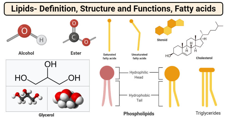
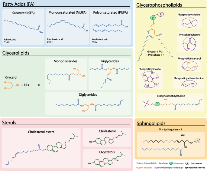
Lipid family overview: fatty acids are building blocks for several lipid classes, while phospholipids, triacylglycerols and steroids have distinct structures and biological roles.
Lipids are an exceptionally diverse group of biological molecules that are grouped together not by a shared structural monomer (like all other macromolecules), but by a shared physical property: they are entirely or almost entirely hydrophobic (insoluble in aqueous solutions like water or blood). However, they dissolve readily in non-polar organic solvents like ethanol, chloroform, or ether. They contain Carbon, Hydrogen, and Oxygen, but with a drastically lower proportion of Oxygen atoms compared to carbohydrates, making them highly reduced.
Figure 6: The vast structural diversity of the Lipid family. This umbrella term includes everything from simple linear fatty acids to bulky energy-storing triglycerides, complex amphipathic membrane phospholipids, and the heavy 4-ring structures of steroid hormones.
The Fundamental Building Blocks: Fatty Acids
A fatty acid consists of a highly acidic, polar, hydrophilic carboxyl functional group ($-COOH$) attached firmly to a long, completely non-polar, hydrophobic hydrocarbon chain (the R-group tail). The length of this hydrocarbon tail (typically 12-24 carbons) and the specific presence or absence of double bonds definitively determine the physical properties (like melting point) of the fat.
Fatty Acid Type
Chemical Structure ($C-C$ bonds)
Physical Properties
Primary Dietary Sources
Saturated
Contains absolutely zero double bonds between carbon atoms. Every single carbon is fully "saturated" with the maximum theoretically possible number of covalently bonded Hydrogen atoms.
Solid (at room temp $20^\circ C$) Straight chains pack tightly together, maximizing strong intermolecular Van der Waals forces.
Molecular Geometry: Saturated vs. Unsaturated Architectures
Clinical Nutrition & Biochemistry
Essential Fatty Acids & The Danger of Trans Fats
Omega ($\omega$) Nomenclature
Mammals lack the specific desaturase enzymes required to introduce double bonds beyond carbon-9 in a fatty acid chain. Therefore, certain highly polyunsaturated fats must be consumed entirely through diet. These are termed "Essential".
The "Omega" number simply indicates the position of the very first double bond, counting backwards starting from the methyl ($CH_3$) end (the omega end) of the chain, rather than the carboxyl ($COOH$) alpha end.
Omega-3 ($\alpha$-linolenic acid): Highly anti-inflammatory precursors. Critical for brain/retina development and synthesizing anti-inflammatory eicosanoids. Sources: Fatty fish (Salmon), flaxseeds, walnuts.
Omega-6 (Linoleic acid): Precursors to pro-inflammatory eicosanoids (prostaglandins, leukotrienes). Essential, but massive overconsumption in modern western diets drives chronic systemic inflammation. Sources: Cheap vegetable seed oils.
The Pathology of Trans Fats
Trans fats are overwhelmingly artificial, created by the industrial food process of partial hydrogenation (bombarding cheap, liquid vegetable oils with hydrogen gas at extreme high heat to make them solid and shelf-stable, like margarine and shortening).
This violent process chemically flips the natural cis double bonds into unnatural trans double bonds. A trans double bond removes the kink! It physically straightens the hydrocarbon chain, forcing an unsaturated fat to behave physically exactly like a saturated fat (solid at room temp).
Cardiovascular Pathology: Human lipase enzymes cannot properly digest trans fats. They radically increase LDL (bad cholesterol), plummet HDL (good cholesterol), and trigger massive systemic inflammation and rapid atherosclerosis (arterial plaque buildup). They are effectively toxic.
🧈 Saturated FA
No C=C double bonds. Straight chains pack efficiently, raising melting point. Commonly solid at room temperature.
🫒 Cis-unsaturated FA
Cis double bonds introduce kinks, reduce packing and lower melting point. They increase membrane fluidity.
⚠️ Trans FA
Trans double bonds keep chains relatively straight and behave more like saturated fats. Industrial trans fats are associated with adverse lipoprotein profiles.
Ω Omega notation
The omega number counts from the methyl end to the first double bond. Humans cannot introduce double bonds beyond Δ9.
🌱 Essential FA
Linoleic acid (ω-6) and α-linolenic acid (ω-3) must be supplied by diet and serve as precursors for longer PUFA pathways.
🔥 Why fats store more energy
Fatty-acid carbons are highly reduced and stored with little water, so oxidation yields much more usable energy per gram than hydrated carbohydrate stores.
🥁6. Triglycerides (Triacylglycerols)
IMAT Challenge
Question 6 Challenge
2022 - Q25
Three fatty acids with the formula C8H16O2 react with glycerol (C3H8O3) to form a triglyceride. Which of the following is the correct formula for the triglyceride?
Triglycerides are the primary, most highly concentrated form of long-term energy storage in the biological world, found in the adipose tissue of animals and the seeds/oils of plants. Because they are entirely non-polar and hydrophobic, they are stored in a completely anhydrous (water-free) state inside cells, making them incredibly lightweight and space-efficient compared to water-logged glycogen.
Biochemical Formation (Esterification)
A triglyceride is synthesized via a massive triple-condensation reaction between exactly 1 molecule of Glycerol (a 3-carbon alcohol backbone) and 3 molecules of Fatty Acids. This reaction forms three covalent Ester Bonds. Because it is a 3-part condensation, the synthesis of one single triglyceride molecule forcibly releases exactly 3 molecules of Water ($H_2O$).
Saponification (Soap Making)
Saponification is the alkaline hydrolysis of the ester bonds in a triglyceride. When a triglyceride is boiled with a strong base (like $NaOH$ or $KOH$), the ester bonds are cleaved, releasing free glycerol and the sodium/potassium salts of the fatty acids, which we universally call Soap. These amphipathic soap molecules can surround dirt and oil, forming micelles that wash away in water.
Figure 7: The chemical skeletal structure of a triglyceride. The vertical backbone on the far left is derived from glycerol. The three long horizontal chains extending to the right are the fatty acid tails, attached via the oxygen atoms of the newly formed ester linkages.
Yields 9 kcal/g (38 kJ/g), more than double the energy density of carbohydrates or proteins. Because it perfectly repels water, it takes up vastly less physical volume and weight in an organism compared to heavily hydrated glycogen.
🧁 Thermal Insulation
Adipose tissue acts as a superb thermal insulator against severe cold. Marine mammals (whales, seals, walruses) rely heavily on extremely thick layers of subcutaneous fat called blubber to survive in freezing ocean temperatures.
🛡 Mechanical Protection
Visceral fat deposits form protective, shock-absorbing cushions around vital internal organs, most notably the delicate kidneys and the heart, protecting them from severe physical trauma and impact.
💧 Metabolic Water Source
The complete aerobic oxidation of lipid hydrocarbon chains in the mitochondria yields a massive amount of $H_2O$ as a byproduct. Desert animals (like the Camel's hump) store immense amounts of fat specifically to be burned to generate vital metabolic water during severe droughts.
🔗 Esterification
Glycerol has three hydroxyl groups; each can react with a fatty-acid carboxyl group. Three condensation reactions form three ester bonds and release three water molecules.
🧼 Saponification
Strong-base hydrolysis of ester bonds produces glycerol plus fatty-acid salts (soap). This is the chemical reverse of ester formation under strongly alkaline conditions.
🌊 7. Phospholipids, Sphingolipids & Membranes
IMAT Challenge
Question 18 Challenge
2013 - Q35
The diagram below shows a phospholipid. When this molecule is broken into a phosphate, glycerol and two fatty acids, the number of water molecules released is:
P (Phosphate)
GLYCEROL
Fatty acid
Fatty acid
Phospholipid structure: The charged phosphate-containing head is hydrophilic, whereas the two hydrocarbon tails are hydrophobic. In water this amphipathic geometry drives spontaneous bilayer formation, with heads facing the aqueous phases and tails sheltered inside.
Phospholipids (specifically glycerophospholipids) are the absolute fundamental architectural basis of all biological membranes on Earth. They are created when one of the three fatty acid tails of a standard triglyceride is removed and replaced by a highly negatively charged, bulky Phosphate Group. This singular chemical substitution completely revolutionizes the physics of the molecule, transforming it from a simple, entirely hydrophobic fat droplet into a highly sophisticated, two-faced Amphipathic molecule.
The "Head" Region: Composed of the glycerol backbone, the phosphate group, and often an additional polar organic molecule attached to the phosphate (like choline, ethanolamine, or serine). Because the phosphate carries a heavy negative charge, the head is extremely polar and fiercely Hydrophilic (Water-loving).
The "Tail" Region: Composed of the two remaining long hydrocarbon fatty acid chains. These chains are completely non-polar, possess zero electrical charge, cannot form hydrogen bonds, and are therefore fiercely Hydrophobic (Water-fearing). Usually, one tail is straight (saturated) and the other is kinked (unsaturated), which precisely dictates the physical fluidity of the membrane.
Sphingolipids: The Specialized Membrane Lipids
Not all membrane lipids use a glycerol backbone. Sphingolipids use a complex amino alcohol called sphingosine as their backbone. The most famous example is Sphingomyelin, which is heavily concentrated in the Myelin Sheath that wraps around and electrically insulates the axons of neurons in the central and peripheral nervous systems, allowing for saltatory conduction of action potentials.
Clinical Note:Multiple Sclerosis (MS) is an autoimmune disease where the immune system destroys this sphingomyelin-rich sheath, crippling nerve transmission. Additionally, a genetic defect in the lysosomal breakdown of sphingolipids leads to catastrophic accumulation diseases, such as Tay-Sachs disease.
When millions of these amphipathic molecules are dumped into an aqueous environment (like the watery interior and exterior of a human cell), the absolute laws of physics and thermodynamics force them to spontaneously self-assemble. To perfectly hide their hydrophobic tails from the water while simultaneously exposing their hydrophilic heads to the water, they instantly form a highly stable, self-sealing double layer known as a Phospholipid Bilayer. This dynamic bilayer is the matrix of the Fluid Mosaic Model of cell membranes.
Architectural Schematic: The Phospholipid Bilayer
A highly detailed representation of a cell membrane. Note how the phospholipid bilayer creates an impenetrable hydrophobic core. This oily core strictly prevents the free passage of water, ions, and large polar molecules, necessitating the existence of complex embedded transport proteins to act as selectively permeable gates.
Clinical Biochemistry
Lipoproteins: The Taxis of the Bloodstream
Blood is overwhelmingly composed of water. Lipids (triglycerides and cholesterol) are strictly hydrophobic; if dumped directly into the blood from the liver or intestines, they would instantly coalesce into massive, lethal fat embolisms that would clog arteries and stop the heart. To safely transport lipids through the aqueous circulatory system, the body packages them into highly complex, water-soluble spheres called Lipoproteins.
A lipoprotein is structured like a biochemical submarine: It features an outer monolayer composed of amphipathic phospholipids and special identifying structural proteins called Apolipoproteins (the hydrophilic heads face the blood). The hollow interior core is stuffed entirely full of highly hydrophobic triglyceride cargo and cholesterol esters.
LDL (Low Density Lipoprotein)
Clinically termed the "Bad Cholesterol". LDL acts as a delivery truck, transporting cholesterol manufactured in the liver out into the peripheral tissues and blood vessels. If a patient has excess LDL, it aggressively invades the endothelial walls of arteries, undergoes rapid oxidation, and gets eaten by macrophages. These macrophages swell and die, forming a fatty streak that hardens into Atherosclerotic Plaque, massively increasing the risk of myocardial infarctions (heart attacks) and strokes.
HDL (High Density Lipoprotein)
Clinically termed the "Good Cholesterol". HDL acts as the body's vascular garbage truck. It constantly circulates through the bloodstream, physically scavenging excess, rogue cholesterol from the peripheral tissues and artery walls, actively pulling it out of developing plaques. It safely transports this dangerous cholesterol back to the liver, where it is metabolized into bile acids and permanently excreted from the body via the intestines (Reverse Cholesterol Transport).
🧠 Sphingolipids
Built on a sphingosine backbone rather than glycerol. Sphingomyelin is abundant in myelin; glycosphingolipids participate in cell recognition.
🩸 Glycolipids
Carbohydrate-bearing lipids are concentrated on the extracellular leaflet and contribute to the glycocalyx and antigenic identity.
🫧 Micelle vs bilayer
Single-tail amphiphiles often favor micelles; two-tail phospholipids are more cylindrical and favor bilayers and closed liposomes.
💍 8. Steroids & Waxes
IMAT Challenge
Question 7 Challenge
2022 - Q26
Which of the following is correct for cholesterol in humans?
Despite having absolutely zero physical or structural resemblance to triglycerides or phospholipids (they contain no fatty acid chains whatsoever), Steroids are formally classified as lipids exclusively because they are fiercely hydrophobic and completely insoluble in water.
Figure 9: The universal, highly distinctive structural fingerprint of all steroids: three 6-sided cyclohexane rings (A, B, C) perfectly fused to one 5-sided cyclopentane ring (D). This rigid planar structure is formally known as the cyclopentanoperhydrophenanthrene nucleus.
Steroids in Human Biology
All steroid hormones in the human body are synthesized directly from a single master precursor molecule: Cholesterol. Because steroid hormones are uniquely lipid-soluble, they do not bind to receptors on the outside of target cells like bulky peptide hormones do. Instead, they slip effortlessly straight through the hydrophobic plasma membrane, entering the cytoplasm or nucleus directly to alter gene transcription at the DNA level.
Cholesterol (The Membrane Buffer): Synthesized primarily in the liver by the enzyme HMG-CoA Reductase (the enzyme targeted by Statin drugs to lower cholesterol). Besides making hormones, it physically wedges itself between the phospholipids of animal cell membranes. It acts as a bidirectional "fluidity buffer": at high temperatures, it physically restricts phospholipid movement, preventing the membrane from melting. At freezing temperatures, its bulky structure forces the phospholipids apart, preventing them from packing tightly and freezing solid.
Corticosteroids: Synthesized by the adrenal cortex. Includes Cortisol (the primary long-term stress hormone, which is heavily immunosuppressive and stimulates gluconeogenesis) and Aldosterone (regulates blood pressure by controlling sodium reabsorption in the kidneys).
Sex Hormones: Synthesized primarily by the gonads. Includes Testosterone, Estrogen, and Progesterone. Responsible for sexual differentiation, secondary sex characteristics, and regulating the menstrual cycle and pregnancy.
Waxes
Waxes are highly complex, extremely stable esters formed by the condensation of an incredibly long-chain fatty acid with an equally long-chain complex alcohol. Because their hydrocarbon chains are so massive, they are the most fiercely hydrophobic biological molecules in existence, solid at room temperature.
Biological Role: They are utilized almost exclusively for heavy-duty waterproofing and protection. Plants secrete dense layers of wax over their leaves (the cuticle) to prevent lethal water loss via transpiration in the hot sun. Bees use beeswax to construct the intricate structural architecture of their honeycombs. Birds and waterfowl coat their feathers in wax secreted from a preen gland to remain buoyant and dry.
Steroid nucleus: the characteristic four fused carbon rings provide the rigid core shared by cholesterol and steroid hormones.
🧱 Membrane cholesterol
Cholesterol fits between phospholipids and buffers membrane fluidity while reducing permeability to small polar molecules.
🧪 Steroid hormones
Glucocorticoids, mineralocorticoids and sex steroids derive from cholesterol and signal mainly through intracellular receptors that regulate transcription.
☀️ Vitamin D
A cholesterol-derived secosteroid pathway links skin UV exposure with calcium/phosphate homeostasis after activation in liver and kidney.
🟡 Bile acids
Cholesterol is converted to amphipathic bile acids, which emulsify dietary lipids and assist formation of mixed micelles in the intestine.
📨 Eicosanoids
Local lipid mediators such as prostaglandins, thromboxanes and leukotrienes are produced from 20-carbon polyunsaturated fatty-acid precursors.
🛡️ Waxes
Long-chain fatty-acid esters of long-chain alcohols form highly hydrophobic protective coatings on skin, feathers and plant surfaces.
🧪 9. Analytical Biochemistry: Lab Tests for Lipids
Testing for lipids is fundamentally different from testing for carbohydrates or proteins. Because lipids lack reactive functional groups (they are essentially just inert hydrocarbon chains), we cannot use a simple colorimetric chemical reaction (like Benedict's or Biuret). Instead, the standard laboratory test for lipids relies entirely on their defining physical property: their strict insolubility in water.
The Emulsion Test
Physical Chemistry Theory: Lipids are completely non-polar, meaning they cannot dissolve in polar water. However, they dissolve very easily in non-polar or moderately polar organic solvents, such as pure absolute Ethanol. By forcing the lipid to dissolve in ethanol first, and then violently shocking the solution by pouring it into a large volume of cold water, the lipid molecules are forcefully ripped out of solution. Because they cannot interact with the water, they instantly precipitate, clumping together to form millions of microscopic, insoluble fat droplets suspended in the water. This suspension of droplets is called an emulsion.
Strict Methodology:
Take a solid or liquid food sample and place it in a completely dry, grease-free test tube.
Add 5ml of absolute, concentrated Ethanol to the tube.
Shake the tube violently for 1-2 minutes. (This physically extracts and fully dissolves any lipids present in the sample into the ethanol).
Decant (pour) the liquid portion containing the dissolved lipid into a second, clean test tube filled with cold, distilled Water.
Positive Result: The immediate formation of a thick, cloudy, opaque White Emulsion near the top of the water.
Physics Note: The emulsion appears milky white due to the Tyndall effect—the millions of tiny suspended lipid droplets physically scatter the light hitting the tube.
Visual Result
Positive: Cloudy White Emulsion
⚡ 10. Thermodynamics: Energy Yield & Respiratory Quotient
A frequent, high-yield IMAT question asks to compare the physical and chemical properties of carbohydrates versus lipids as energy storage molecules. Why does a mobile animal store the vast bulk of its energy as fat rather than just building a massive brick of glycogen?
Biochemical Feature
Carbohydrates (Glycogen)
Lipids (Triglycerides)
Physiological Use Case
Short-Term, Rapid Energy.(Burned during sprinting or immediate stress. Soluble, easy to mobilize).
Long-Term, Dense Energy.(Burned during resting, endurance, or starvation).
Total Energy Density
~17 kJ/g (4 kcal/g)
~38 kJ/g (9 kcal/g)
Deep Chemistry Reason
Carbohydrates are already partially oxidized. They contain a massive amount of heavy Oxygen atoms ($-OH$ groups) relative to Carbon. Because they are already partially oxidized, completing their combustion yields significantly less net energy.
Lipids are highly, heavily reduced. They consist almost entirely of non-polar C-H bonds with virtually zero oxygen atoms. Oxidizing these pure hydrocarbon chains in the mitochondria yields vast quantities of high-energy electrons for the ETC.
Physical Storage Weight
Carbohydrates are intensely hydrophilic. In cells, 1 gram of glycogen chemically binds to and traps about 2-3 grams of heavy Water. Storing massive amounts of energy as glycogen would make an animal far too heavy and bulky to walk or escape predators.
Lipids are fiercely hydrophobic. They are stored completely anhydrously (without a single drop of water) in dense fat droplets inside adipocytes. This extreme weight efficiency is an absolute evolutionary mandate for mobile animals and flying birds.
The Respiratory Quotient (RQ)
The RQ is a dimensionless number used in basal metabolic rate calculations to determine exactly which macronutrient a patient or organism is currently burning for energy. It is calculated by measuring the volume of $CO_2$ exhaled divided by the volume of $O_2$ consumed during respiration over time.
$RQ = \frac{\text{Volume of } CO_2 \text{ produced}}{\text{Volume of } O_2 \text{ consumed}}$
RQ = 1.0 (Carbohydrates): Glucose oxidation uses 6 $O_2$ to produce 6 $CO_2$. ($6/6 = 1$).
RQ = ~0.7 (Lipids): Because lipids have so little oxygen inherently, they require a massive intake of $O_2$ from the air to be fully oxidized into $CO_2$ and $H_2O$. Thus, the denominator ($O_2$ consumed) is much larger.
RQ = ~0.9 (Proteins): An intermediate value.
Essential IMAT Glossary
Anomeric Carbon: The new chiral carbon atom formed entirely upon the ring closure of a linear sugar. It is C1 in an aldose (glucose) and C2 in a ketose (fructose). It is the chemically reactive center of the molecule.
Amphipathic: A unique structural property of a molecule possessing both a fiercely hydrophilic, polar region and a fiercely hydrophobic, non-polar region. Phospholipids and cholesterol are classic examples.
Glycosidic Bond: The specific type of covalent bond formed via a condensation reaction that uniquely links two monosaccharides together to form disaccharides or polysaccharides.
Ester Bond: The specific covalent bond formed during a condensation reaction between the highly acidic carboxyl group of a fatty acid and the hydroxyl (alcohol) group of glycerol, assembling triglycerides.
Chirality (Stereocenter): An absolute geometric property of some molecules. A carbon atom is chiral if it is covalently bonded to four completely different atoms or functional groups, making the molecule entirely non-superimposable on its mirror image (enantiomer), drastically affecting its interaction with specific biological enzymes.
📝 Mastery Practice: IMAT Biomolecules Quiz
Test your comprehensive understanding of the advanced carbohydrate and lipid biochemistry, molecular structures, and clinical lab analytics covered in this module. These questions are meticulously styled after the rigorous, integrated, multi-step logic required for the IMAT examination.