Lesson 7: Complete Histology
A comprehensive histology atlas: tissue architecture, microscopic recognition, staining logic, structure–function relationships, and IMAT-focused distinctions.
Lesson 7: Complete Histology
Read tissue from structure to function: epithelial architecture → extracellular matrix → neural organization → muscle microanatomy. The goal is to identify what you actually see in histological preparations and explain why the structure fits the function.
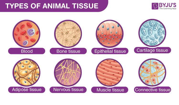
Histology is not memorizing photographs. A reliable diagnosis begins with scale → stain → architecture → dominant cell type → extracellular material → special surface → vascular pattern. Use this sequence every time you face an unfamiliar micrograph.
🔎 0.1 A six-question slide-reading algorithm
🎨 0.2 H&E and other stains — why the colors look the way they do
| Stain / method | Main target | Typical color | What becomes easy to see | Classic use |
|---|---|---|---|---|
| Hematoxylin | Acidic structures: DNA, RNA, ribosomes | Blue-purple | Nuclei, nucleoli, RER-rich cytoplasm | Routine H&E; basophilia |
| Eosin | Basic/eosinophilic proteins | Pink-red | Collagen, cytoplasmic proteins, muscle | Routine H&E; eosinophilia |
| PAS | Carbohydrate-rich molecules | Magenta | Basement membrane, glycogen, mucins | Kidney, liver glycogen, fungal walls |
| Masson trichrome | Collagen vs muscle | Collagen blue/green; muscle red | Fibrosis and connective-tissue organization | Scar, myocardium, liver fibrosis |
| Alcian blue | Acidic mucopolysaccharides / GAGs | Blue | Mucin and cartilage ground substance | Goblet cells, cartilage |
| Silver stain | Reticular fibers / selected neural structures | Black-brown | Type III collagen networks | Lymphoid organs, liver stroma |
| Oil Red O / Sudan | Neutral lipid in frozen sections | Red-orange | Lipid droplets | Adipose, fatty change |
| Immunohistochemistry | Specific antigen | Chromogen-dependent | Cell lineage / protein expression | Keratin, vimentin, GFAP, desmin, CD markers |
🧪 0.3 Processing artifacts and microscopy
| Method | Strength | Limitation | Histology clue |
|---|---|---|---|
| Light microscopy | Whole-tissue architecture and routine staining | Resolution ~0.2 μm | Best for H&E pattern recognition |
| TEM | Internal ultrastructure | Thin sections, complex preparation | Basal lamina, junctions, organelles, sarcomeres |
| SEM | Surface topography | Does not show internal section detail | Microvilli, cilia, surface architecture |
| Frozen section | Rapid, preserves lipid and enzyme activity better | Lower morphology quality | Used intraoperatively and for lipid stains |
| Paraffin section | Excellent routine morphology | Lipids extracted; shrinkage artifacts possible | Most standard teaching slides |
Histology and microscopy
Apply this section immediately with official IMAT past-paper questions. Select an answer to reveal the full worked explanation.
The diagrams show five different microscopic structures. The structures are not drawn to the same scale. Which of these structures is the smallest?
| Structure | Magnification / Scale bar given |
|---|---|
| 1 | Drawing length: 4cm, Mag: ×4000 |
| 2 | Max diameter: 3cm, Mag: ×400 |
| 3 | Scale bar: 0.14 mm |
| 4 | Scale bar: 0.5 μm |
| 5 | Drawing length: 6cm, Mag: ×20000 |
The decisive statement is 4
This question tests Histology and microscopy. Apply the definitions, structural relationships, and cause-and-effect rules in the section immediately above; the remaining choices conflict with at least one of those conditions.
Test every condition in the stem independently. Reject an option as soon as one statement contradicts the biological rules established in this lesson.
Which sequence shows cells of increasing size (from left to right)?
The decisive statement is E. coli → human red blood cell → onion epidermal cell
This question tests Histology and microscopy. Apply the definitions, structural relationships, and cause-and-effect rules in the section immediately above; the remaining choices conflict with at least one of those conditions.
Test every condition in the stem independently. Reject an option as soon as one statement contradicts the biological rules established in this lesson.
An electron microscope (EM) was used to view a cell. The maximum dimension of the cell was observed at a magnification ×30000. The image on the EM screen showed a maximum length of 30 mm. Which of the following mature healthy cells was being magnified?
The decisive statement is one coccus bacterium in a Staphylococcus cluster
This question tests Histology and microscopy. Apply the definitions, structural relationships, and cause-and-effect rules in the section immediately above; the remaining choices conflict with at least one of those conditions.
Test every condition in the stem independently. Reject an option as soon as one statement contradicts the biological rules established in this lesson.
Epithelial Tissue
Epithelium is a continuous sheet of cells that covers body surfaces, lines cavities, and forms glands. It is avascular, supported by a basement membrane, and exhibits polarity.
2.1 Classification & Location
| Layers | Shape | Type | Key Locations & Functions |
|---|---|---|---|
| Simple (1 Layer) | Squamous | Simple Squamous | Alveoli, Endothelium, Mesothelium. (Rapid Diffusion/Filtration) |
| Cuboidal | Simple Cuboidal | Kidney Tubules, Thyroid Follicles. (Secretion/Absorption) | |
| Columnar | Simple Columnar | Tall polarized cells specialized for secretion or absorption; apical microvilli may be present. | |
| Stratified (>1 Layer) | Squamous | Stratified Squamous | Keratinized: Epidermis (Dry, Waterproof). Non-keratinized: Oral cavity, Vagina (Moist). (Protection) |
| Columnar | Pseudostratified | Respiratory Tract (Trachea). Ciliated. (Mucus Transport) | |
| Variable | Transitional | Bladder, Ureter. Dome cells. (Stretch/Distension) |
Visual: Classification of Epithelial Tissues and Cell Polarity

How to read: Epithelial Classification & Polarity
Classification Grid (Left)
A complete visual guide to epithelial types based on layer count and cell shape:
- Layers: Simple (1 layer), Stratified (>1 layer), and Pseudostratified.
- Shapes: Squamous (flat), Cuboidal (cube), Columnar (tall), and Transitional (variable).
- Context: Examples for each type (e.g., Alveoli for Simple Squamous, Trachea for Ciliated Pseudostratified) are integrated.
Cell Polarity & Junctions (Right)
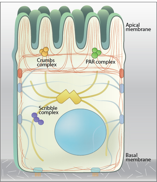
A high-magnification view of a single epithelial cell detailing its functional domains:
- Apical Domain: Features like microvilli (absorption) and cilia (transport).
- Lateral Domain: The "junctional complex" (Tight, Adherens, Desmosomes, Gap) with molecular details like claudins, cadherins, and connexons.
- Basal Domain: Anchoring to the basement membrane (Basal Lamina + Reticular Lamina) via Hemidesmosomes (integrins).
2.2 Surface Specializations — What You Actually See
| Specialization | Core structure | Histological meaning |
|---|---|---|
| Microvilli | Actin bundles | Increase apical surface area; individually below light-microscope resolution, collectively form a brush/striated border. |
| Motile cilia | 9+2 microtubule axoneme + dynein | Longer apical projections that move luminal material. |
| Primary cilium | 9+0 microtubule axoneme | Usually one per cell; sensory/signaling organelle rather than a motile brush. |
| Keratin layer | Intermediate-filament-rich dead surface cells | Strong barrier against abrasion and water loss. |
Epithelial tissue
Apply this section immediately with official IMAT past-paper questions. Select an answer to reveal the full worked explanation.
Which of the following is/are tissues?
1 cartilage
2 skin
3 endothelium
3. Endothelium: This is a specialized, single-cell-thick layer of epithelial tissue that lines the interior surface of blood vessels.
Squamous epithelium, ciliated epithelium and cartilage are tissues found in healthy humans. Which one of the following structures is made up of at least TWO of these tissues?
Capillaries: Made only of a single layer of squamous endothelium.
Bowman's capsule: Made of specialized squamous epithelium (podocytes).
Oviduct (Fallopian tube): Contains ciliated epithelium to move the egg, but lacks cartilage (it uses smooth muscle instead).
2.3 Polarity, Junctions, Basement Membrane & Tissue Renewal
Histology should be read from cell architecture, not from memorized organ stories. First identify layers and apical shape, then ask how polarity, junctions and the basement membrane create the tissue's barrier and mechanical behavior.
Histology recognition sequence
Layer count and apical shape define the epithelial name
| Pattern | Microscopic clue | Primary mechanical / transport logic |
|---|---|---|
| Simple squamous | very thin flattened cells | rapid diffusion / filtration |
| Simple cuboidal | round central nuclei; cell height ≈ width | secretion / controlled transport |
| Simple columnar | tall polarized cells; basal nuclei | secretion / absorption |
| Stratified squamous | many layers; superficial cells flattened | resists abrasion |
| Pseudostratified | nuclei at different heights; all cells basal | specialized secretion / surface transport |
| Transitional | rounded dome cells when relaxed | reversible distension without barrier failure |
An epithelial cell is three functional membrane domains in one cell
Do not memorize junction names without their mechanical partner
| Junction | Core proteins | Cytoskeletal link | Primary function |
|---|---|---|---|
| Tight junction | Claudins, occludin | Actin via ZO proteins | Seals paracellular route; preserves apical/basolateral polarity |
| Adherens junction | Classical cadherins | Actin via catenins | Adhesion belt; tissue shape and contractile tension |
| Desmosome | Desmoglein, desmocollin | Intermediate filaments | High mechanical strength between neighboring cells |
| Gap junction | Connexins → connexons | no load-bearing linkage | Direct ion / small-molecule communication |
| Hemidesmosome | Integrin α6β4 | Keratin IF | Anchors epithelial cell to basal lamina |
| Focal adhesion | Integrins, talin, vinculin | Actin | Dynamic cell-ECM adhesion and mechanosensing |
The basement membrane is an organized signaling and mechanical platform
Epithelia are dynamic populations, not static sheets
self-renewal maintains the compartment
transit-amplifying cells expand rapidly
polarity and tissue-specific proteins emerge
apoptosis or shedding is balanced by renewal
- Contact inhibition: intact epithelial sheets restrain unnecessary proliferation.
- Loss of basement membrane: disrupts polarity and survival signaling; many epithelial cells undergo anoikis when detached.
- EMT: epithelial cells can lose E-cadherin/polarity and gain migratory mesenchymal traits during development, repair and cancer invasion.
- Regeneration: basal lamina often acts as a scaffold guiding restoration after superficial injury.
Classify glands by architecture and secretion mechanism
| Question | Categories | What the microscope shows |
|---|---|---|
| Duct present? | Exocrine vs endocrine | Exocrine glands retain ducts; endocrine cells release to interstitium/capillaries. |
| Duct branching? | Simple vs compound | Simple = unbranched duct; compound = branched duct system. |
| Secretory-unit shape? | Tubular vs acinar/alveolar | Elongated tube vs rounded secretory end-piece. |
| Secretion appearance? | Serous vs mucous | Serous cells stain darker with round nuclei; mucous cells appear pale/foamy with flattened basal nuclei. |
| Release mechanism? | Merocrine / apocrine / holocrine | Exocytosis / apical cytoplasm loss / whole-cell disintegration. |
IMAT histology rule: classify first, then connect junctions and matrix to mechanics. Pseudostratified epithelium is one true layer; type IV collagen and laminin belong to the basal lamina; desmosomes link intermediate filaments, while adherens junctions link actin.
Embryological Origin & Core Properties
Epithelium can arise from all three germ layers: ectoderm produces epidermis and corneal epithelium; mesoderm produces endothelium and mesothelium; endoderm produces much of the gastrointestinal and respiratory lining. What unites these tissues is not embryonic origin but architecture: high cellularity, little extracellular matrix, apical–basal polarity, attachment to basement membrane, avascularity, and rapid renewal in many sites.
Classification: two questions only
First count true layers; then name the shape of the most apical cells. Pseudostratified epithelium is one layer because every cell contacts the basement membrane even though nuclei lie at different heights. Transitional epithelium is stratified and changes superficial-cell shape during distension.

| Type | Microscopic clue | Typical role | Representative site |
|---|---|---|---|
| Simple squamous | One extremely thin layer; flattened nuclei | Diffusion / filtration / lubrication | Alveoli, endothelium, mesothelium |
| Simple cuboidal | Round central nuclei; height ≈ width | Absorption / secretion | Kidney tubules, thyroid follicles |
| Simple columnar | Tall cells; basal oval nuclei | Absorption / secretion | GI lining, gallbladder |
| Pseudostratified columnar | Nuclei at multiple heights; all cells basal | Secretion / surface transport | Respiratory epithelium |
| Stratified squamous | Many layers; flat apical cells | Abrasion resistance | Skin, oral cavity, esophagus |
| Transitional | Dome-shaped umbrella cells when relaxed | Stretch without barrier failure | Ureter, bladder |
Histological examples — read the epithelium, not the organ story


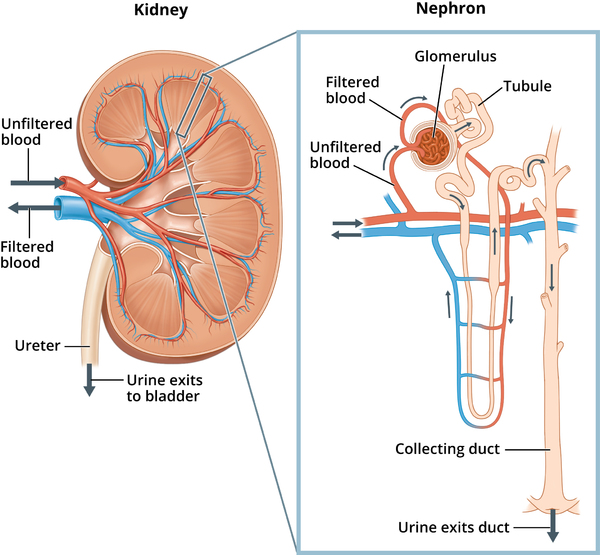
Stomach mucosa: surface protection and deep glandular secretion in the same epithelial sheet
The gastric surface is lined by simple columnar mucous cells that produce a bicarbonate-rich protective mucus. The epithelium invaginates into gastric pits, which lead into glands. In the fundus/body, parietal cells are large and eosinophilic because they are mitochondria-rich and secrete HCl plus intrinsic factor; chief cells are more basophilic because their abundant rough ER synthesizes pepsinogen. Mucous neck cells and enteroendocrine cells complete the glandular population.

Liver lobule: a glandular epithelial organ organized around blood flow and bile flow in opposite directions
Hepatocytes are epithelial cells arranged in one-cell-thick plates radiating toward a central vein. Between plates lie sinusoids, which receive mixed blood from branches of the portal vein and hepatic artery located in portal triads. Bile, however, enters bile canaliculi between adjacent hepatocytes and flows toward bile ductules in the portal triad—roughly opposite to sinusoidal blood flow.

| Structure | What it contains | Direction / role | Recognition clue |
|---|---|---|---|
| Portal triad | Portal venule + hepatic arteriole + bile ductule | Blood enters; bile exits | Three distinct profiles in connective tissue at lobule edge |
| Sinusoid | Mixed portal + arterial blood | Toward central vein | Irregular vascular channels between hepatocyte plates |
| Space of Disse | Perisinusoidal exchange space | Plasma–hepatocyte exchange | Contains stellate (Ito) cells |
| Kupffer cell | Resident macrophage | Phagocytosis | Located along sinusoidal lining |
Basement Membrane & Hemidesmosomes
The basement membrane is more than a support sheet. The basal lamina contains laminin, type IV collagen, nidogen and perlecan; the reticular lamina is contributed by connective tissue. Hemidesmosomes use α6β4 integrin and intracellular linker proteins such as plectin to couple keratin intermediate filaments to laminin-rich matrix. This arrangement lets epithelium resist shear while remaining polarized.
Renewal
Epithelia are frequently renewing tissues. Small-intestinal epithelium turns over in roughly 4–6 days, whereas epidermal replacement is slower. Stem/progenitor cells divide in protected niches, differentiate as they migrate, and are ultimately shed or undergo programmed death.
The key epithelial question is not merely “what shape is the cell?” It is how the sheet is polarized, attached, renewed and specialized for exchange, secretion, absorption or protection.
🧭 E1. Classification logic — layers first, apical shape second
| Pattern | Recognition rule | Function | Examples | Common trap |
|---|---|---|---|---|
| Simple squamous | One layer of very flat cells; flattened nuclei | Diffusion, filtration, lubrication | Alveoli, endothelium, mesothelium | Do not call every thin lining “endothelium”; endothelium specifically lines vessels/heart. |
| Simple cuboidal | Round central nuclei; cell height ≈ width | Absorption / secretion | Renal tubules, thyroid follicles, small ducts | Cross-sections of tubules can mimic circular glands. |
| Simple columnar | Tall cells; oval nuclei usually basal | Absorption / secretion | GI tract, gallbladder | Look for goblet cells and brush border to refine location. |
| Pseudostratified columnar | Nuclei at different heights but every cell contacts basement membrane | Mucus secretion + transport | Respiratory tract | Looks stratified; it is actually one layer. |
| Stratified squamous | Many layers; surface cells are flat | Protection against abrasion | Skin, esophagus, oral cavity, vagina | Name by surface cell shape, not basal cells. |
| Transitional / urothelium | Multilayered with dome-shaped umbrella cells when relaxed | Stretch + urine barrier | Ureter, bladder | Umbrella cells flatten with distension. |
🧬 E2. Polarity and apical specializations
Microvilli
Short, non-motile projections containing an actin core. They increase surface area and are especially dense in absorptive cells. In the small intestine they form the brush/striated border; in proximal renal tubules they produce a fuzzy luminal edge.
Stereocilia
Despite the name, stereocilia are very long actin-based microvilli, not true cilia. They occur in the epididymis and sensory hair cells of the inner ear.
Motile cilia
Built on a 9+2 microtubule axoneme with dynein. Coordinated beating moves mucus in the respiratory tract and the oocyte in the uterine tube. Basal bodies show a centriole-like 9-triplet arrangement.
Primary cilium
Usually a single 9+0 sensory cilium. It acts as a signaling antenna rather than a propulsive organelle and is important in Hedgehog signaling and mechanosensation.
Cell polarity is the organizing principle behind epithelial transport
An epithelial cell is not a symmetrical box. The apical membrane faces a lumen or the exterior, the lateral membrane contacts neighboring cells and contains junctional complexes, and the basal membrane interfaces with the basement membrane and connective tissue. This compartmentalization lets the cell place different transporters on opposite surfaces and therefore move substances directionally across the epithelium.
🔗 E3. Junctional complex — molecules, cytoskeleton and function
| Junction | Core proteins | Cytoskeletal link | Main job | Clinical / exam link |
|---|---|---|---|---|
| Tight junction | Claudins, occludin, ZO proteins | Actin-associated scaffolds | Seals paracellular pathway; preserves polarity | Perineurium and BBB-type barriers depend on tight junctions. |
| Adherens junction | Classical cadherins + catenins | Actin | Mechanical belt; cell-shape remodeling | Ca²⁺-dependent adhesion. |
| Desmosome | Desmoglein, desmocollin | Intermediate filaments | Spot-weld mechanical strength | Prominent in epidermis and myocardium. |
| Gap junction | Connexins → connexons | Not primarily anchoring | Passes ions/small molecules between cells | Electrical coupling in cardiac/smooth muscle. |
| Hemidesmosome | Integrin α6β4, plectin | Intermediate filaments | Anchors cell to basal lamina | Cell–matrix junction, not cell–cell. |
🧪 E4. Basement membrane — what is actually in it?
The basement membrane seen by light microscopy includes a basal lamina produced largely by epithelial cells plus a deeper reticular component contributed by connective tissue. The basal lamina contains type IV collagen, laminin, nidogen and perlecan. It provides anchorage, polarity cues and selective filtration.
🧴 E5. Glandular epithelium — classification beyond “exocrine vs endocrine”
| Feature | Option A | Option B | Histology clue | Examples |
|---|---|---|---|---|
| Destination | Exocrine: secretion enters a duct / surface | Endocrine: secretion enters blood/interstitium | Endocrine glands lack ducts and are highly vascular | Salivary vs thyroid/pituitary |
| Secretory unit | Tubular | Acinar / alveolar | Shape of terminal secretory portion | Intestinal glands vs pancreas |
| Duct system | Simple | Compound | Branched duct tree = compound | Sweat gland vs salivary gland |
| Release mode | Merocrine: exocytosis | Apocrine / holocrine: apical cytoplasm or whole-cell loss | Look for apical blebs or disintegrating cells | Pancreas / mammary lipid / sebaceous |
♻️ E6. Renewal, metaplasia and barrier adaptation
Many epithelia renew continuously from stem/progenitor cells. Intestinal epithelium turns over in only a few days, whereas other epithelia renew more slowly. Chronic stress can induce metaplasia: one differentiated epithelial type is replaced by another better suited to the environment. This adaptation may protect short-term but can increase disease risk if the stress persists.
🧠 E7. Practical recognition atlas
| Specimen | First thing to find | Signature epithelial clue | Supporting clue | Do not confuse with |
|---|---|---|---|---|
| Trachea | Lumen + cartilage | Pseudostratified ciliated columnar + goblet cells | Hyaline cartilage rings, glands | Esophagus |
| Esophagus | Thick mucosa | Non-keratinized stratified squamous | No villi; muscular wall | Skin |
| Small intestine | Villi | Simple columnar + goblet cells + brush border | Crypts | Colon (no villi) |
| Kidney cortex | Glomeruli + tubules | Simple cuboidal tubules | PCT fuzzy lumen vs DCT clearer lumen | Exocrine gland acini |
| Urinary bladder | Large folded lumen | Transitional epithelium with umbrella cells | Thick smooth muscle wall | Stratified squamous mucosa |
| Skin | Keratin layer | Keratinized stratified squamous | Hair follicles / glands / dermis | Oral mucosa |
Connective Tissue
Characterized by sparse cells scattered in an abundant Extracellular Matrix (ECM).
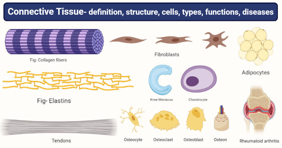
Figure 3.1: Varieties of Connective Tissue
3.1 Extracellular Matrix (ECM)
Protein Fibers
- Collagen Type I: Thick, eosinophilic bundles. High tensile strength. (Bone, Skin, Tendon).
- Collagen Type II: Thin fibrils. (Hyaline Cartilage).
- Collagen Type III (Reticular): Delicate meshwork. (Lymph nodes, Spleen, Liver). Silver stain.
- Elastic Fibers: Elastin core + Fibrillin. Stretch and recoil. (Aorta, Ear).
Ground Substance
A hydrated gel that resists compression.
- GAGs: Long, negative sugar chains (Hyaluronic acid, Chondroitin sulfate). Attract Na⁺ and water.
- Proteoglycans: Core protein + GAGs (Aggrecan).
- Glycoproteins: Adhesive glue (Fibronectin, Laminin).
3.2 Specialized Tissues
Bone & Cartilage
- Avascular. Cells in Lacunae.
- Hyaline: Type II Collagen. (Joints, Ribs).
- Elastic: Elastic fibers. (Ear).
- Fibrocartilage: Type I. (Discs).
- Calcified Matrix (Hydroxyapatite). Vascular.
- Osteoblasts: Build bone.
- Osteoclasts: Resorb bone (Macrophage origin).
- Osteon: Haversian system in compact bone.
Cartilage histology is a matrix problem: lacunae, territorial matrix and the absence of vessels
Cartilage contains chondrocytes in lacunae embedded in a highly hydrated matrix. Because cartilage is avascular, nutrients must diffuse through the matrix. Matrix immediately around lacunae is often darker (territorial matrix) because it is especially rich in sulfated proteoglycans; the interterritorial matrix lies farther away. Hyaline cartilage is type-II-collagen-rich, elastic cartilage adds elastic fibers, and fibrocartilage contains conspicuous type I collagen bundles.

| Type | Fiber emphasis | Perichondrium | Classic sites |
|---|---|---|---|
| Hyaline | Type II collagen, visually “glassy” | Usually present; absent at articular surfaces | Trachea, costal cartilage, joints |
| Elastic | Type II + elastic fibers | Present | Epiglottis, auricle |
| Fibrocartilage | Type I collagen bundles | Absent | IV discs, pubic symphysis, menisci |
Blood
- Erythrocytes (RBC): 7.5µm. Anucleate biconcave discs. Transport O₂. Energy via Anaerobic Glycolysis only.
- Leukocytes (WBC):
Granulocytes: Neutrophil (Bacteria), Eosinophil (Allergy/Parasite), Basophil (Histamine).
Agranulocytes: Lymphocyte (T/B Cells), Monocyte (Macrophage precursor). - Platelets: Thrombocytes. Fragments of Megakaryocytes. Clotting.
Connective tissue
Apply this section immediately with official IMAT past-paper questions. Select an answer to reveal the full worked explanation.
Which of the following are components of the skin?
1 adipose tissue
2 blood capillaries
3 erector muscle
2. Blood capillaries: The dermis is highly vascularized to supply nutrients to the epidermis and aid in thermoregulation (vasodilation/constriction).
3. Erector muscle: The arrector pili are tiny smooth muscles in the dermis attached to hair follicles; they contract to stand hair up (goosebumps) to trap heat. All three are integral structural components.
What is the main role of osteoblasts?
Where in a shoulder joint are osteocytes found?
The decisive statement is bone tissue
This question tests Connective tissue. Apply the definitions, structural relationships, and cause-and-effect rules in the section immediately above; the remaining choices conflict with at least one of those conditions.
Test every condition in the stem independently. Reject an option as soon as one statement contradicts the biological rules established in this lesson.
3.3 Cell–Matrix Adhesion, Mechanotransduction & ECM Remodeling
Connective tissue is not inert packing material. Cells continuously build, sense, pull on and remodel their extracellular matrix. Matrix composition determines tensile strength, compression resistance, elasticity and cell behavior.
ECM has a controlled life cycle
fibroblasts secrete collagen, proteoglycans and glycoproteins
fibers align and cross-link outside the cell
MMPs cleave matrix proteins
TIMPs limit MMP activity and prevent excessive tissue destruction
Remodeling is essential for development and wound repair, but excessive protease activity can weaken tissue and facilitate tumor invasion.
Healing changes the matrix over time
| Phase | Dominant cells / signals | Matrix event |
|---|---|---|
| Hemostasis | platelets, fibrin | temporary clot scaffold |
| Inflammation | neutrophils → macrophages | debris clearance; cytokine release |
| Proliferation | fibroblasts, endothelial cells | granulation tissue; collagen III-rich provisional matrix |
| Remodeling | fibroblasts / myofibroblasts | collagen maturation and stronger type I-rich scar |
Mechanics rule: collagen carries tension, hydrated proteoglycans resist compression, elastin permits recoil, and integrins let cells sense the force carried by that matrix.
Connective tissue is defined by its extracellular matrix. The exam-relevant question is usually not “which cells are present?” but “which matrix component gives this tissue its mechanical property?”
Collagen types you should separate instantly
Type I
Bone, tendon, skin, scar. Strong tensile fibers; most abundant collagen.
Type II
Hyaline/elastic cartilage and vitreous body. Resists pressure in cartilage matrix.
Type III
Reticular fibers in lymphoid organs and soft tissues; delicate supporting meshwork.
Type IV
Sheet-forming collagen in basal laminae rather than fibrils; supports epithelial interfaces.
Collagen synthesis — intracellular → extracellular
Cartilage, bone and blood — same tissue family, different matrix
| Tissue | Dominant matrix logic | Cells | High-yield clue |
|---|---|---|---|
| Cartilage | Hydrated proteoglycan matrix + type II collagen | Chondrocytes in lacunae | Avascular; heals slowly |
| Bone | Type I collagen + hydroxyapatite | Osteoblast / osteocyte / osteoclast | Mineralized and vascular |
| Blood | Fluid matrix = plasma | RBCs, WBCs, platelets | Fibers appear during clotting as fibrin |
| Adipose | Sparse ECM; energy-rich cellular tissue | Adipocytes | Endocrine organ as well as storage |
IMAT pattern: type IV collagen belongs to basal lamina; type II points to cartilage; type I points to tendon/bone/skin; type III forms reticular networks.
Connective tissue is defined by the dominance of extracellular matrix rather than tightly packed cells. The matrix contains collagen, elastic and reticular fibers suspended in hydrated ground substance rich in GAGs, proteoglycans and adhesive glycoproteins.
Adipose Tissue
| Feature | White adipose | Brown adipose |
|---|---|---|
| Lipid droplets | One large unilocular droplet | Many small multilocular droplets |
| Nucleus | Flattened and peripheral | More central |
| Mitochondria | Relatively fewer | Very abundant; UCP1/thermogenin |
| Major role | Energy storage, cushioning, endocrine signaling | Non-shivering thermogenesis |
Adipose is an endocrine connective tissue, not merely an inert fat store
White adipocytes contain a single large lipid droplet, a thin rim of cytoplasm and a flattened peripheral nucleus. They store triacylglycerol, cushion organs and secrete adipokines such as leptin and adiponectin. Brown adipocytes contain many lipid droplets and abundant mitochondria; UCP1 dissipates the proton gradient as heat rather than coupling it fully to ATP synthesis.

Cartilage
| Type | Dominant fibers | Perichondrium | Key clue |
|---|---|---|---|
| Hyaline | Type II collagen + aggrecan | Usually present; absent at articular surface | Glassy matrix; most common cartilage |
| Elastic | Type II collagen + elastic fibers | Present | Flexible framework |
| Fibrocartilage | Thick type I collagen | Absent | Rows of chondrocytes between dense collagen bundles |
Bone Cells & Osteon
Compact bone is organized into osteons (Haversian systems): concentric lamellae surround a central vascular canal. Osteocytes occupy lacunae and extend processes through canaliculi; Volkmann canals connect adjacent osteons and periosteal vessels.
Bone cell lineage: builder, embedded sensor, and resorptive giant
Osteoblasts arise from mesenchymal osteoprogenitors and secrete osteoid, chiefly type I collagen plus matrix proteins. When an osteoblast becomes enclosed by matrix it differentiates into an osteocyte, which occupies a lacuna and sends processes through canaliculi to sense mechanical load. Osteoclasts come from the monocyte/macrophage lineage, are multinucleated, attach to bone at a sealing zone and resorb mineralized matrix using acid plus proteases.
Ossification & Growth Plate
Intramembranous ossification forms bone directly from mesenchyme. Endochondral ossification replaces a cartilage model. In the epiphyseal plate, learn the sequence: resting → proliferation → hypertrophy → calcification → ossification.
Intramembranous and endochondral ossification reach bone by different routes
Intramembranous ossification forms bone directly within a mesenchymal membrane and is important for flat skull bones and part of the clavicle. Endochondral ossification first creates a hyaline cartilage model, then replaces that model with bone. Longitudinal growth at the epiphyseal plate is therefore a highly ordered cartilage-to-bone transition.
| Growth-plate zone | Dominant event | What you see | Memory cue |
|---|---|---|---|
| Resting | Reserve chondrocytes | Scattered small cells | “Reserve” |
| Proliferation | Mitosis | Columns / stacks | “Palisade” |
| Hypertrophy | Cell enlargement | Large lacunae | “Huge” |
| Calcification | Matrix calcifies, cells die | Thin calcified spicules | “Calcified scaffold” |
| Ossification | Osteoblast deposition | New bone on cartilage remnants | “Bone replaces cartilage” |
Blood & Lymph as Connective Tissues
Blood has a fluid matrix (plasma) containing erythrocytes, leukocytes and platelets. Lymphoid tissue provides reticular stromal networks that organize lymphocytes and antigen-presenting cells. The key histology distinction is whether the matrix is fluid, fibrous, mineralized or gel-like.
Blood is connective tissue with a fluid extracellular matrix
In blood, plasma is the extracellular matrix and cells circulate freely. Mammalian erythrocytes are biconcave and anucleate; loss of the nucleus and most organelles creates space for hemoglobin and prevents the cell from consuming the oxygen it transports. Leukocytes retain nuclei and can be classified by nuclear shape, granules and abundance. Platelets are anucleate fragments derived from megakaryocytes.

Lymph node microanatomy turns lymph flow into organized immune surveillance
A lymph node is wrapped by a connective-tissue capsule. Afferent lymphatics deliver lymph to the subcapsular sinus. The superficial cortex contains B-cell follicles, the deeper paracortex is T-cell-rich and contains high endothelial venules, and the medulla contains medullary cords and sinuses. Lymph eventually exits through an efferent vessel at the hilum.

Connective tissue is recognized by the fact that the extracellular matrix often occupies more space than the cells. To read it correctly, separate three variables: cell population, fiber system and ground substance.
🧱 C1. Connective tissue proper — resident and wandering cells
| Cell | Origin / identity | Main function | Histological clue | High-yield link |
|---|---|---|---|---|
| Fibroblast | Mesenchymal stromal cell | Synthesizes collagen, elastin, proteoglycans, glycoproteins | Spindle nucleus between fibers | Activated fibroblasts drive fibrosis and wound repair. |
| Macrophage | Monocyte lineage | Phagocytosis, antigen presentation, cytokines | Irregular outline; lysosome-rich cytoplasm | Resident macrophages have tissue-specific names. |
| Mast cell | Hematopoietic | Histamine, heparin, inflammatory mediators | Granule-rich cell near vessels | Immediate hypersensitivity. |
| Plasma cell | Activated B-cell lineage | Antibody secretion | Eccentric “clock-face” nucleus, basophilic cytoplasm | RER-rich antibody factory. |
| Adipocyte | Mesenchymal | Lipid storage / endocrine function | White fat: signet-ring appearance | Leptin, adiponectin; brown fat uses UCP1. |
🧵 C2. Fibers — molecular composition predicts mechanics
| Fiber | Molecule | Mechanical role | Where prominent | Stain / clue |
|---|---|---|---|---|
| Collagen I | Thick collagen fibrils | Tensile strength | Tendon, dermis, bone | Eosinophilic; trichrome highlights collagen |
| Collagen II | Fine fibrils in proteoglycan-rich matrix | Compression-resistant framework | Hyaline and elastic cartilage | Fibrils often not individually visible in H&E |
| Collagen III | Reticular fibers | Delicate supporting meshwork | Lymphoid organs, liver, marrow | Silver-staining / argyrophilic |
| Collagen IV | Sheet-forming collagen | Basal-lamina network | Basement membranes | PAS-positive context |
| Elastic fibers | Elastin + fibrillin | Recoil after stretch | Elastic arteries, lung, elastic ligaments | Special elastic stains improve visibility |
💧 C3. Ground substance — the hydrated molecular gel
Ground substance contains GAGs, proteoglycans, adhesive glycoproteins and tissue fluid. Negative charges on GAGs attract Na⁺ and water, creating a hydrated gel that resists compression and permits diffusion. Hyaluronan is unusual because it is not sulfated and is synthesized at the plasma membrane rather than in the Golgi.
🪢 C4. Loose vs dense connective tissue
| Type | Fiber arrangement | Cells / matrix | Mechanical behavior | Typical location |
|---|---|---|---|---|
| Loose areolar | Loosely arranged mixed fibers | Many cells + abundant ground substance | Flexible packing / diffusion | Lamina propria under epithelia |
| Dense regular | Parallel collagen bundles | Few fibroblasts, little ground substance | Strong in one axis | Tendon, ligament |
| Dense irregular | Interwoven collagen bundles | Fibroblasts between bundles | Resists multidirectional tension | Dermis, organ capsules |
| Reticular tissue | Type III collagen mesh | Supports many free cells | Soft stromal framework | Lymph node, spleen, marrow |
🟡 C5. Adipose tissue — white vs brown
| Feature | White adipose | Brown adipose | Why it matters |
|---|---|---|---|
| Lipid droplets | One large unilocular droplet | Multiple small multilocular droplets | Changes cell shape and nuclear position |
| Nucleus | Flattened at periphery | More central | Brown fat does not look like a simple signet ring |
| Mitochondria | Moderate | Very abundant | UCP1 uncouples oxidation from ATP synthesis to produce heat |
| Vascularity | Good | Very rich | Supports thermogenesis |
🧊 C6. Cartilage — cells, matrix and growth
Cartilage is avascular; chondrocytes receive nutrients by diffusion through matrix. Chondroblasts secrete matrix and become trapped in lacunae as chondrocytes. Most cartilage has a perichondrium, but articular hyaline cartilage and fibrocartilage are important exceptions.
| Type | Major fibers | Appearance | Location | Special point |
|---|---|---|---|---|
| Hyaline | Type II collagen | Glassy basophilic matrix; isogenous groups | Trachea, costal cartilage, articular surfaces | Most common; articular cartilage lacks perichondrium |
| Elastic | Type II + elastic fibers | Chondrocytes in lacunae plus dark elastic network | External ear, epiglottis | Flexible and resilient |
| Fibrocartilage | Type I dominant | Rows of chondrocytes between thick collagen bundles | IV discs, pubic symphysis, menisci | No perichondrium; strongest tensile component |
🦴 C7. Bone histology — matrix, osteon and remodeling
Bone matrix combines an organic osteoid phase (mainly type I collagen) with inorganic hydroxyapatite. This composite design resists both tension and compression. Osteoblasts build matrix, osteocytes maintain it through canaliculi, and multinucleated osteoclasts resorb it.
| Cell | Origin | Main action | Histology | Molecular cue |
|---|---|---|---|---|
| Osteoblast | Mesenchymal osteoprogenitor | Secretes osteoid; mineralization | Cuboidal row on bone surface | RANKL production helps regulate osteoclastogenesis |
| Osteocyte | Entrapped osteoblast | Mechanosensing and matrix maintenance | Cell in lacuna with canaliculi | Communicates through gap junctions |
| Osteoclast | Monocyte/macrophage lineage | Bone resorption | Large multinucleated cell in Howship lacuna | Ruffled border + H⁺ pumps + lysosomal enzymes |
Compact bone is organized into osteons with concentric lamellae around Haversian canals; Volkmann canals connect vascular channels. Spongy bone is organized into trabeculae and lacks classic osteons in many regions.
🩸 C8. Blood and hematopoietic tissue
| Cell | Nucleus | Key morphology | Main function | Recognition clue |
|---|---|---|---|---|
| Erythrocyte | Absent in mature mammalian RBC | Biconcave eosinophilic disc | Gas transport | Central pallor; no nucleus |
| Neutrophil | 3–5 lobes | Fine pale granules | Acute bacterial defense | Segmented nucleus |
| Eosinophil | Bilobed | Large red-orange granules | Parasites / allergy | Very eosinophilic granules |
| Basophil | Often obscured | Dark blue-purple granules | Histamine-rich inflammatory response | Rare; granules cover nucleus |
| Lymphocyte | Large round | Thin rim of cytoplasm | Adaptive immunity | High nucleus:cytoplasm ratio |
| Monocyte | Kidney/horse-shoe | Abundant gray-blue cytoplasm | Macrophage precursor | Largest common circulating leukocyte |
🛡️ C9. Lymphoid histology — node, spleen and thymus
| Organ | Architecture | Signature structure | Main traffic / job | High-yield distinction |
|---|---|---|---|---|
| Lymph node | Cortex + paracortex + medulla | Follicles; medullary cords/sinuses | Filters lymph | Has afferent lymphatics |
| Spleen | White pulp + red pulp | Central arterioles, sinusoids | Filters blood | No cortex/medulla organization like lymph node |
| Thymus | Lobules with dark cortex + pale medulla | Hassall corpuscles | T-cell maturation | No lymphoid follicles in normal thymus |
🩹 C10. Wound repair and fibrosis
Muscle Tissue
Muscle tissue is specialized for contraction via the interaction of Actin and Myosin filaments. It consumes ATP to generate force.
4.1 Classification
| Type | Striations | Control | Structure | Features |
|---|---|---|---|---|
| Skeletal | Yes | Voluntary | Cylindrical, Multinucleated (peripheral) | Triads (T-tubule + 2 SR). Troponin C. |
| Cardiac | Yes | Involuntary | Branched, 1-2 Nuclei (central) | Intercalated Discs (Gap junctions + Desmosomes). Diads. |
| Smooth | No | Involuntary | Fusiform, 1 Nucleus (central) | Dense Bodies (no sarcomeres). Calmodulin (no Troponin). |
4.2 The Sarcomere & Contraction
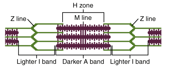
Band Changes
- A Band: Length of myosin. Remains constant.
- I Band: Actin only. Shortens.
- H Zone: Myosin only. Shortens.
- Z Lines: Move closer together.
Excitation-Contraction Coupling
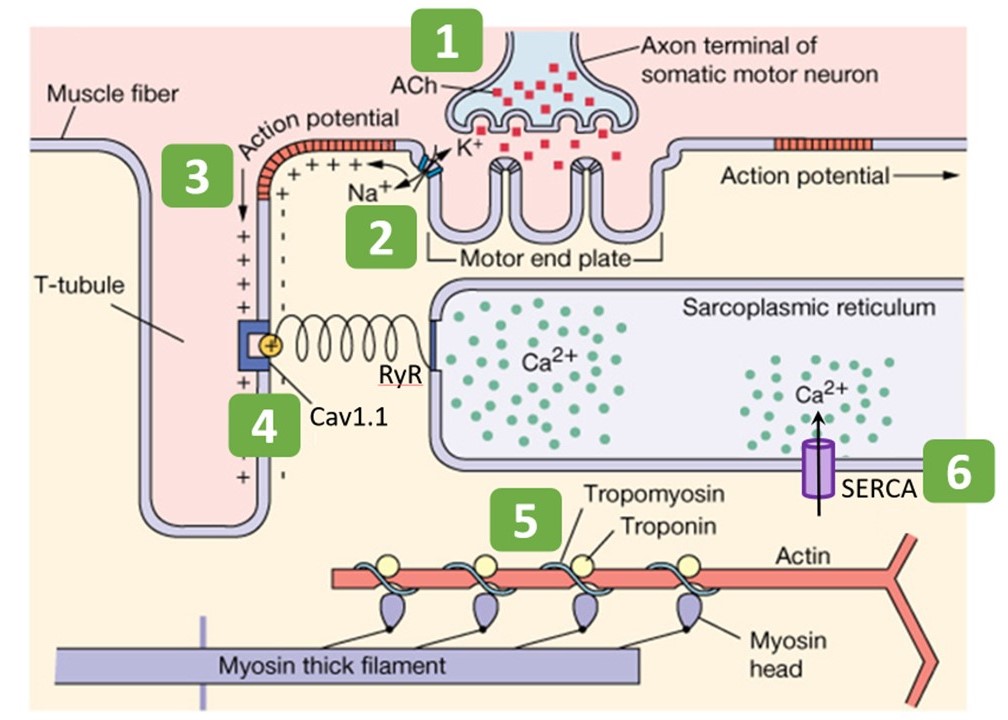
- Nerve AP releases ACh -> Muscle AP.
- AP travels down T-tubules to Triad.
- Voltage sensor (DHP) opens Ryanodine Receptors (RyR) on SR.
- Ca²⁺ floods sarcoplasm.
- Ca²⁺ binds Troponin C.
- Troponin moves Tropomyosin off actin sites.
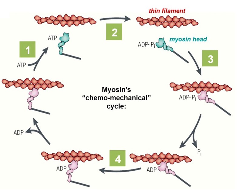
- Myosin heads bind actin (Crossbridge) → Power Stroke (ATP needed for release).
4.3 From Excitation to Force
Keep one causal chain in mind: membrane excitation → Ca²⁺ release → regulatory proteins move → cross-bridges cycle → sarcomere shortens → ATP restores the system.
Histology first: skeletal vs cardiac vs smooth

Intercalated discs combine mechanical and electrical coupling

Follow the hierarchy before memorizing the sarcomere


Only overlap changes; filament length does not

The membrane signal is converted into a Ca²⁺ signal
Force comes from repeated cross-bridge cycling


4.4 ATP Supply & Fiber-Type Strategy
After the contraction mechanism is clear, the remaining high-yield question is how muscle sustains ATP demand and how fiber types trade speed for endurance.
| ATP source | Speed | Capacity | Best use |
|---|---|---|---|
| Stored ATP | Immediate | Very low | First seconds |
| Creatine phosphate | Very fast | Low | Short explosive effort |
| Anaerobic glycolysis | Fast | Moderate | High-intensity short duration |
| Oxidative phosphorylation | Slower | High | Endurance |
Three Muscle Types
| Feature | Skeletal | Cardiac | Smooth |
|---|---|---|---|
| Striations | Present | Present | Absent |
| Cells | Long cylindrical fibers | Short branched cells | Fusiform cells |
| Nuclei | Many, peripheral | Usually one central; sometimes two | One central |
| Control | Voluntary somatic motor | Involuntary; intrinsic rhythm + autonomic modulation | Involuntary; autonomic, hormonal, local |
| Special hallmark | Triads, motor end plates | Intercalated discs, dyads | Dense bodies, caveolae, no troponin |
Skeletal Muscle Hierarchy
Epimysium surrounds the whole muscle; perimysium surrounds fascicles; endomysium surrounds individual muscle fibers. These connective-tissue layers merge into tendons and provide a mechanical path from microscopic sarcomere shortening to macroscopic movement.
Sarcomere & Excitation–Contraction Coupling
The sarcomere extends from Z disc to Z disc. Thin filaments are anchored at Z discs; thick filaments center around the M line. During contraction, the A band stays essentially constant while the I band and H zone shorten. In skeletal muscle, an action potential enters T-tubules, activates voltage-sensing DHPR, opens RyR1 in the sarcoplasmic reticulum, raises cytosolic Ca²⁺, and allows Ca²⁺ to bind troponin C.
Clinical / Functional Correlates
Rigor mortis occurs because ATP depletion prevents myosin detachment from actin. Duchenne muscular dystrophy results from dystrophin deficiency, destabilizing the sarcolemma during contraction. A motor unit is one α-motor neuron plus all muscle fibers it innervates; small units permit fine control, while large units generate powerful coarse movements.
Cardiac Muscle
Cardiomyocytes are branched, striated and joined by intercalated discs. Fascia adherens and desmosomes transmit mechanical force; gap junctions permit electrical coupling. Cardiac excitation–contraction coupling relies on Ca²⁺ entry through L-type channels followed by calcium-induced calcium release from RyR2 channels.
Smooth Muscle
Smooth muscle lacks sarcomeres and troponin. Actin anchors to dense bodies. Ca²⁺ binds calmodulin, activating MLCK, which phosphorylates myosin light chains and increases myosin ATPase activity. The latch state permits prolonged tension with low ATP consumption.
Muscle is easiest to identify by three questions: Are there striations? Where are the nuclei? Are cells separate or fused into long fibers? Then add junctions, connective-tissue investment and Ca²⁺-handling structures.
🏗️ M1. Skeletal muscle hierarchy
⚡ M2. Sarcomere landmarks and what changes during contraction
| Region | Contains | During contraction | Reason |
|---|---|---|---|
| A band | Full length of thick filaments | Length stays constant | Myosin filaments do not shorten |
| I band | Thin filaments only | Shortens | Actin slides toward M line |
| H zone | Thick filaments only | Shortens / may disappear | Overlap increases |
| Z disc | Thin-filament anchoring boundary | Z discs move closer | Sarcomere shortens |
Skeletal muscle converts a membrane action potential into a rapid Ca²⁺ pulse
The action potential travels along the sarcolemma and down the T-tubule. Voltage-sensitive dihydropyridine receptors (DHPR/CaV1.1) mechanically activate ryanodine receptor 1 in the sarcoplasmic reticulum, releasing Ca²⁺. Ca²⁺ binds troponin C, shifts tropomyosin and allows actin–myosin cross-bridge cycling. SERCA pumps return Ca²⁺ to the SR during relaxation.
🧪 M3. Fiber types — histochemical and metabolic differences
| Feature | Type I | Type IIa | Type IIx / fast glycolytic | Functional consequence |
|---|---|---|---|---|
| Contraction | Slow | Fast | Very fast | Force-speed profile |
| Mitochondria | Many | Intermediate-high | Fewer | Oxidative endurance |
| Myoglobin | High | Intermediate | Low | Red vs pale appearance |
| Capillaries | Dense | Intermediate | Lower | O₂ delivery |
| Fatigue | Resistant | Moderate | Rapid | Posture vs bursts of power |
🧠 M4. Neuromuscular junction and proprioceptors
At the neuromuscular junction, the motor axon terminal sits over a specialized postsynaptic membrane with deep junctional folds enriched in nicotinic ACh receptors. Acetylcholinesterase in the basal lamina rapidly terminates the signal.
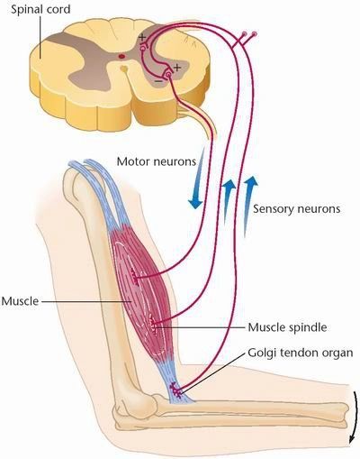
| Receptor | Where | What it senses | Afferent output | Reflex role |
|---|---|---|---|---|
| Muscle spindle | Within muscle, parallel to extrafusal fibers | Muscle length and rate of stretch | Group Ia/II | Stretch reflex; tone |
| Golgi tendon organ | Tendon, in series with fibers | Tension / force | Group Ib | Force feedback |
Muscle spindles measure muscle length; Golgi tendon organs measure tension
A muscle spindle contains intrafusal fibers enclosed within a capsule and arranged in parallel with ordinary extrafusal fibers. Ia and II sensory afferents report length and rate of stretch. Gamma motor neurons adjust intrafusal-fiber tension so the spindle remains sensitive as the muscle changes length. The stretch reflex uses this signal to resist sudden lengthening.
❤️ M5. Cardiac muscle — intercalated discs and energy demand
Cardiomyocytes are branched, striated cells with central nuclei. They are joined end-to-end by intercalated discs, where fascia adherens and desmosomes transmit force while gap junctions spread depolarization. Cardiac cells contain abundant mitochondria because contraction is continuous and heavily aerobic.
| Feature | Skeletal | Cardiac | Smooth | Histology key |
|---|---|---|---|---|
| Nuclei | Many peripheral | 1–2 central | 1 central | Fastest discriminator |
| Striations | Yes | Yes | No | Sarcomeres present in skeletal/cardiac |
| Cell shape | Long cylindrical | Branched | Spindle-shaped | Cardiac branching is characteristic |
| Cell coupling | Fibers electrically separate | Gap junctions at intercalated discs | Often gap-junction coupled in unitary smooth muscle | Electrical syncytium differs by tissue |
| Regeneration | Limited via satellite cells | Very poor | Relatively good | Explains fibrosis after myocardial injury |
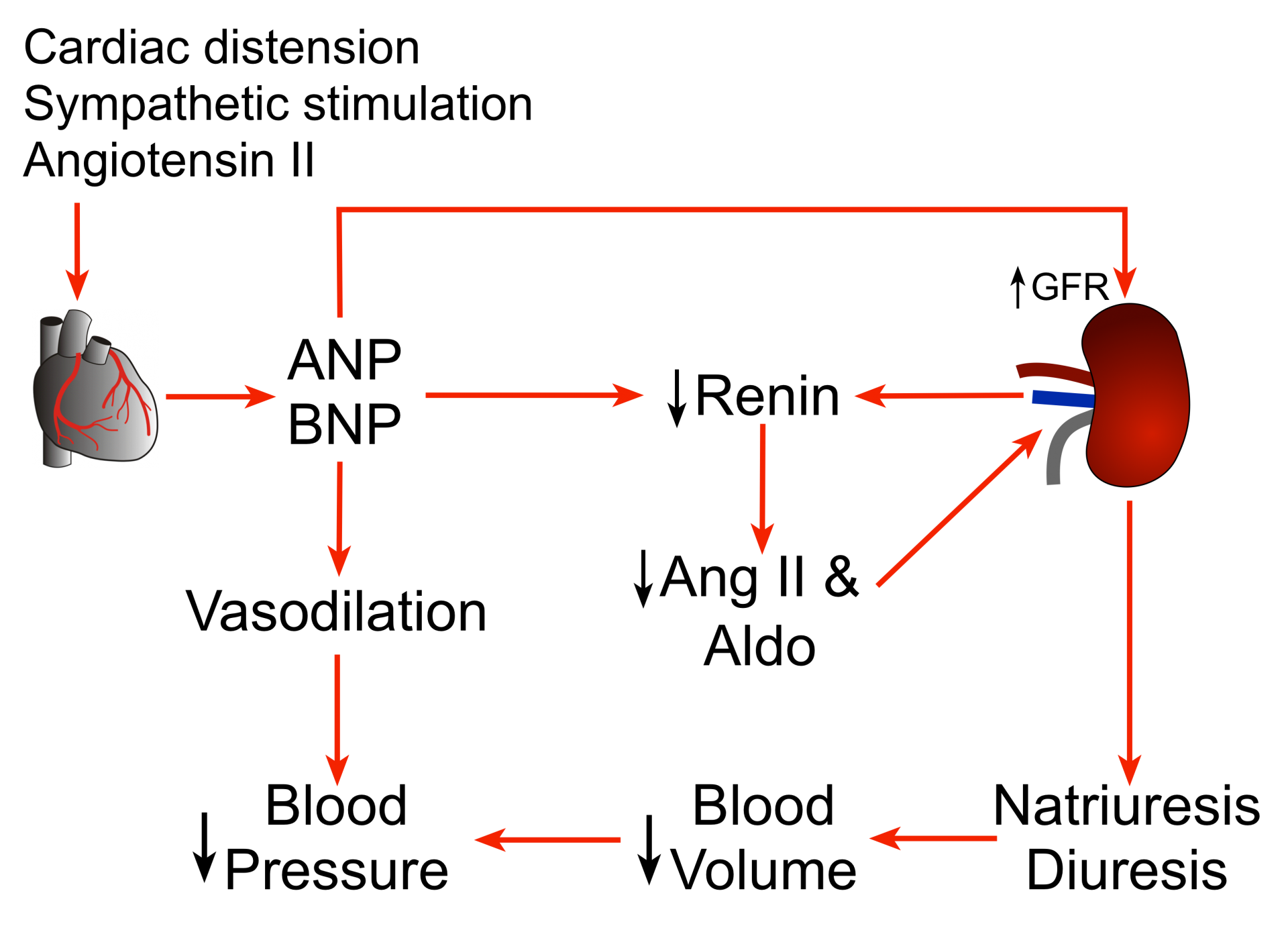
Atrial cardiomyocytes are contractile cells with an endocrine role
Stretch of atrial cardiomyocytes promotes release of atrial natriuretic peptide (ANP). ANP promotes natriuresis, increases renal Na⁺ and water excretion, opposes the renin–angiotensin–aldosterone system and tends to reduce blood volume and pressure. Histologically, atrial myocytes can contain secretory granules in addition to their contractile apparatus.
Intercalated discs combine mechanical strength with low-resistance electrical coupling
Cardiomyocytes must remain physically attached during forceful repetitive contraction while also propagating depolarization rapidly from cell to cell. Fascia adherens anchors actin and transmits contractile force, desmosomes anchor intermediate filaments and resist separation, and gap junctions allow ions to pass directly between cells.

🌊 M6. Smooth muscle — dense bodies, caveolae and plasticity
Smooth muscle cells are spindle-shaped and lack sarcomeres. Actin and intermediate filaments anchor to dense bodies, which function mechanically like dispersed Z-line equivalents. Caveolae help organize membrane Ca²⁺ signaling because smooth muscle lacks a classical T-tubule system.
Unlike skeletal muscle, smooth muscle regulates myosin: Ca²⁺ binds calmodulin, which activates MLCK, phosphorylating the myosin light chain. Smooth muscle can undergo both hypertrophy and hyperplasia.
Smooth muscle regulates myosin rather than using troponin
Ca²⁺ binds calmodulin, and the Ca²⁺–calmodulin complex activates myosin light-chain kinase (MLCK). MLCK phosphorylates the regulatory light chain of myosin and increases myosin ATPase activity. Myosin light-chain phosphatase reverses phosphorylation. Smooth muscle can maintain force even after myosin is dephosphorylated while still attached to actin—the latch state—which saves ATP during sustained tone.
🔬 M7. Slide recognition — fast practical checklist
| Tissue | Longitudinal section | Cross section | Best clue | Common trap |
|---|---|---|---|---|
| Skeletal | Parallel fibers, striations, peripheral nuclei | Large polygonal fibers, peripheral nuclei | Multinucleated syncytial fibers | Dense regular tendon beside muscle |
| Cardiac | Branched fibers, central nuclei, intercalated discs | Variable profiles, central nuclei | Branching + central nuclei | Skeletal muscle if discs are not obvious |
| Smooth | Spindle cells, cigar-shaped central nuclei, no striations | Small variable circular profiles | Sheets of eosinophilic cells without striation | Dense connective tissue |
Nervous Tissue
Nervous tissue allows for rapid communication and integration. It consists of excitable Neurons and supporting Neuroglia.
5.1 Physiology: The Action Potential
The fundamental unit of neural communication.
- Resting (-70mV): Established by Na⁺/K⁺ pump and K⁺ leak channels. Inside is negative.
- Depolarization: Threshold reached. Voltage-gated Na+ Channels open. Na⁺ rushes IN. Potential shoots to +30mV.
- Repolarization: Na⁺ channels inactivate. Voltage-gated K+ Channels open. K⁺ rushes OUT. Potential drops.
- Hyperpolarization: K⁺ channels constitute slow closing. Potential dips below resting level.
- Refractory Period: Ensures one-way propagation.
5.2 Synaptic Transmission
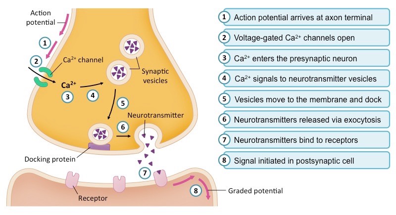
- Action potential arrives at the Axon Terminal.
- Depolarization opens Voltage-gated Ca²⁺ Channels.
- Ca²⁺ influx triggers synaptic vesicles (SNARE proteins) to fuse with membrane.
- Neurotransmitter (NT) is released into the cleft (Exocytosis).
- NT binds to Ligand-gated ion channels on postsynaptic cell.
- Channels open -> Ion flux -> EPSP or IPSP.
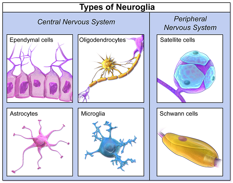
5.3 Neuroglia (Support Cells)
CNS Glia
- Astrocytes: Blood-Brain Barrier, Metabolic support, K+ buffering.
- Oligodendrocytes: Myelinate multiple axons.
- Microglia: Immune defense (Phagocytes).
- Ependymal Cells: Line ventricles, produce CSF.
PNS Glia
- Schwann Cells: Myelinate a single axon segment. Aid regeneration.
- Satellite Cells: Support neuron cell bodies in ganglia.
5.4 Neuron → Channel → Myelin → Synapse
Neurophysiology becomes coherent when you keep four layers separate: ion gradients maintain readiness, voltage-gated channels generate the action potential, myelin accelerates propagation, and synapses convert the signal back into a graded input.
Input, trigger, conduction and output happen in different places


The Na⁺/K⁺ ATPase maintains gradients; channels create millisecond voltage changes
Read the curve as a sequence of channel states
CNS and PNS use different myelinating cells

Ca²⁺ entry is the trigger for vesicle fusion
The initial segment sums graded inputs before deciding to fire

Nervous histology is a pattern-recognition problem. Distinguish neuronal somata vs neuropil, gray vs white matter, CNS vs PNS support cells, and ganglion vs nerve before thinking about electrical physiology.
🧬 N1. Neuron soma — what Nissl substance tells you
Neuronal somata often have a large pale euchromatic nucleus with a prominent nucleolus, reflecting intense transcription. Nissl substance represents RER and free ribosomes and is therefore basophilic. It is present in the soma and proximal dendrites but absent from the axon hillock and axon.
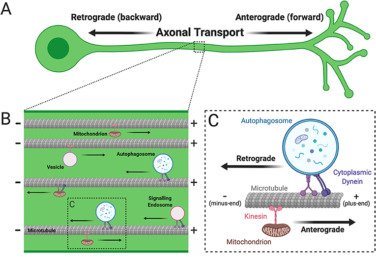
Axons depend on microtubule tracks because their terminals may be far from the soma
Most axonal proteins are synthesized in the soma, not at the distant axon terminal. Microtubules therefore act as polarized tracks: kinesin generally drives anterograde cargo toward the plus end and terminal, whereas dynein carries retrograde cargo toward the soma. Fast transport moves vesicles, mitochondria and membrane components; slower transport moves cytoskeletal proteins and soluble enzymes.
🧩 N2. CNS glia — same tissue, different jobs
| Glial cell | Main job | Histology / marker concept | High-yield distinction |
|---|---|---|---|
| Astrocyte | K⁺ buffering, transmitter uptake, metabolic support, BBB support, scar | Processes often better seen with GFAP IHC than routine H&E | Does not form the endothelial tight junction itself; supports BBB function. |
| Oligodendrocyte | CNS myelin | Small dark round nucleus; limited visible cytoplasm | One cell can myelinate segments of multiple axons. |
| Microglia | Resident immune / phagocytic cell | Small elongated dark nucleus | Mesoderm/yolk-sac lineage rather than neuroectodermal glia. |
| Ependymal cell | Lines ventricles and central canal | Cuboidal/columnar ciliated lining | Specialized ependymal cells contribute to choroid plexus. |
Glial cells create the environment in which neurons can function
Astrocytes regulate extracellular K⁺, recycle neurotransmitters, support metabolism and contribute to the blood–brain barrier through end-feet signaling. Oligodendrocytes form CNS myelin. Microglia are immune phagocytes of yolk-sac/mesodermal lineage. Ependymal cells line ventricles and the central canal; specialized ependymal cells participate in the choroid plexus.
🧵 N3. CNS regional histology
| Region | Organization | Signature cell / layer | What to look for | Common trap |
|---|---|---|---|---|
| Cerebral cortex | Gray matter superficial; white matter deep | Pyramidal neurons | Layered cortex over pale white matter | Do not expect the 3-layer cerebellar pattern |
| Cerebellar cortex | Molecular → Purkinje → granular | Large Purkinje cells in single row | Pale molecular layer + dark granular layer | Purkinje layer is one-cell-thick, not a broad zone |
| Spinal cord | Central gray matter, peripheral white matter | Large motor neurons in ventral horn | Butterfly/H-shaped gray matter | Opposite gray/white arrangement from cerebral cortex |
Gray matter and white matter reverse their relative positions between brain and spinal cord
Gray matter contains neuronal cell bodies, dendrites, synapses and abundant neuropil; white matter is dominated by myelinated axons. In the cerebral and cerebellar cortex, gray matter forms a superficial cortex over deeper white matter. In the spinal cord, gray matter is central and butterfly-shaped, while white matter surrounds it.
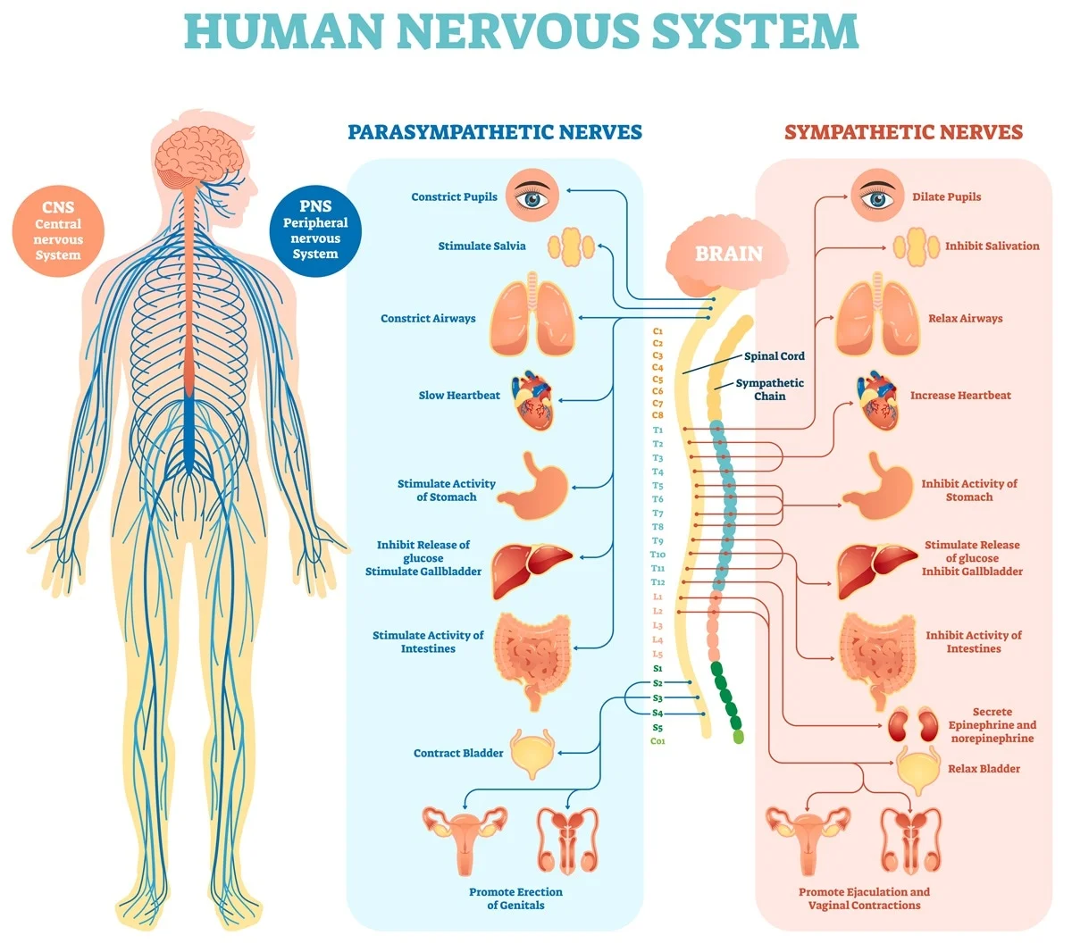
🧷 N4. PNS ganglia — dorsal root vs autonomic
| Feature | Dorsal root ganglion | Autonomic ganglion | Why useful |
|---|---|---|---|
| Neuron type | Pseudounipolar sensory | Multipolar autonomic | Morphology reflects pathway |
| Nucleus | Usually central | Often eccentric | Useful supporting clue |
| Satellite cells | Form a very complete ring around soma | Less regular / less complete | One of the best practical distinctions |
| Synapses in ganglion | No synapse between peripheral and central sensory branch | Preganglionic neurons synapse here | Functional distinction |
🧵 N5. Peripheral nerve coverings and fascicles
Endoneurium surrounds individual nerve fibers, perineurium surrounds fascicles and forms the blood–nerve barrier through tight junctions, and epineurium surrounds the entire nerve and carries larger vessels.
Peripheral nerve coverings are hierarchical and each layer has a different job
Endoneurium surrounds individual nerve fibers, perineurium wraps fascicles and forms a diffusion barrier through tight junctions, and epineurium binds fascicles together and carries larger vessels. A nerve cross-section therefore looks like bundles within bundles.

⚡ N6. Myelin, nodes and degeneration
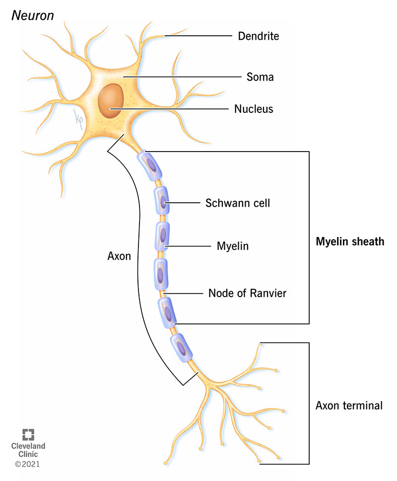
Myelin is a multilayered membrane sheath. In the CNS, oligodendrocytes form myelin; in the PNS, Schwann cells do. Nodes of Ranvier interrupt the sheath and concentrate voltage-gated Na⁺ channels, enabling saltatory conduction.
After PNS axonal injury, distal axon and myelin undergo Wallerian degeneration. Macrophages remove debris, while Schwann cells can form regeneration tubes that guide regrowth if the connective-tissue pathway remains intact. CNS regeneration is much more limited.
Myelin changes membrane physics and creates saltatory conduction
Myelin increases membrane resistance and decreases effective capacitance along internodes, so depolarizing current spreads farther and faster. Voltage-gated Na⁺ channels cluster at nodes of Ranvier, where the action potential is regenerated. In the PNS each Schwann cell forms one internode; in the CNS one oligodendrocyte can myelinate multiple axonal segments.
🧠 N7. Gray matter vs white matter
| Feature | Gray matter | White matter | Microscopic reason |
|---|---|---|---|
| Dominant content | Neuron somata, dendrites, synapses, glia | Myelinated axons + glia | Myelin-rich regions appear paler in routine paraffin H&E |
| Neuropil | Abundant | Less somatic neuropil pattern | Neuropil = interwoven neuronal/glial processes between cell bodies |
| Location example | Cerebral cortex, spinal cord horns | Deep cerebral white matter, peripheral spinal cord | Regional organization differs between brain and spinal cord |
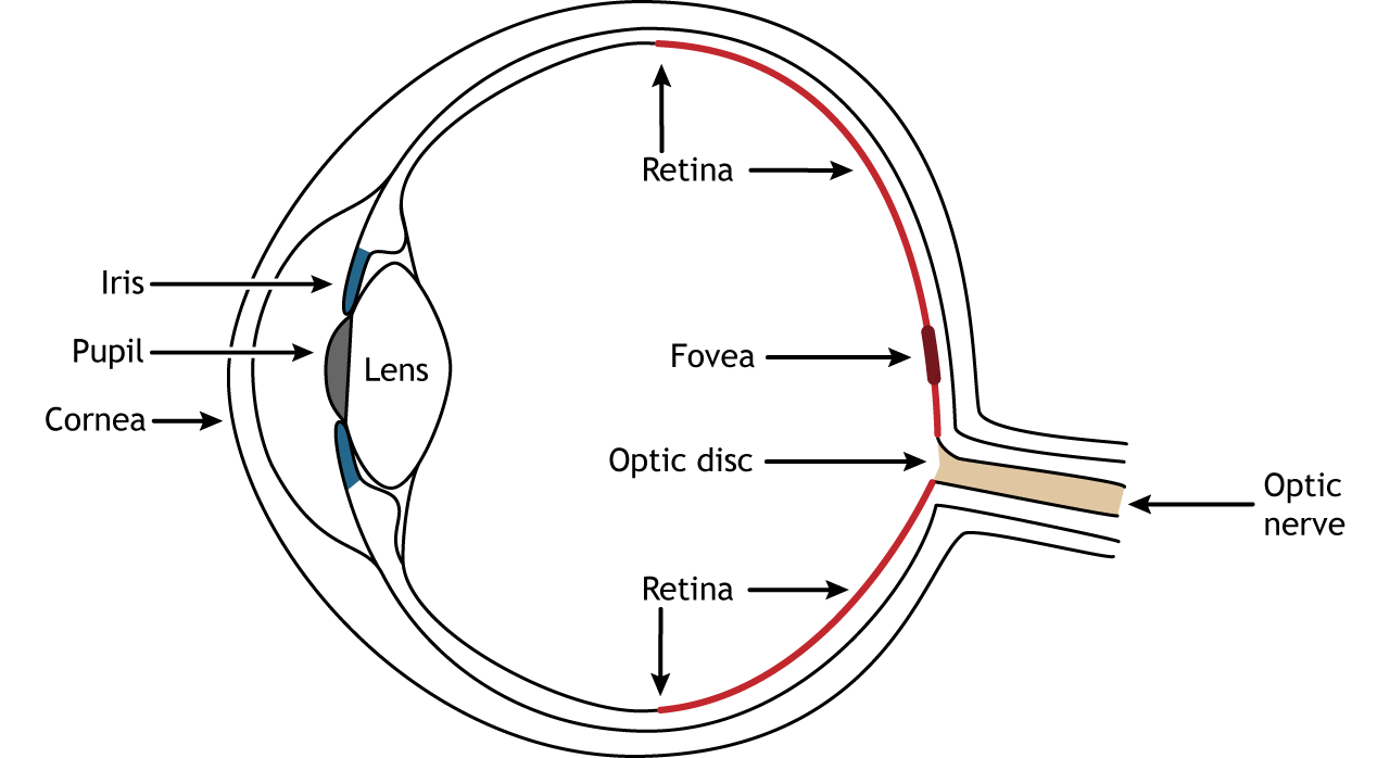
The retina is a layered neural circuit in which light and information travel in opposite directions
Light enters from the vitreous side and passes through ganglion and bipolar cell layers before reaching rods and cones near the retinal pigment epithelium. Neural information then moves in the opposite direction: photoreceptors → bipolar cells → ganglion cells. Horizontal and amacrine cells provide lateral integration. Ganglion-cell axons converge to form the optic nerve.
🩸 N8. Blood–brain barrier — histological logic
The BBB depends on continuous capillary endothelial cells joined by tight junctions, a continuous basal lamina, pericytes and astrocytic end-feet. Astrocytes support barrier properties but the physical paracellular seal is formed by the endothelial junctions.
🔬 N9. Practical neural slide identification
| Specimen | Find first | Signature clue | Supporting clue | Trap |
|---|---|---|---|---|
| Cerebellum | Cortical layering | Purkinje-cell row | Dark granular layer | Cerebrum |
| Spinal cord | Central canal + H-shaped gray | Large ventral motor neurons | Peripheral white matter | Brainstem sections |
| Dorsal root ganglion | Large round neuron somata | Complete satellite-cell rings | Central nuclei | Autonomic ganglion |
| Peripheral nerve | Fascicles | Perineurial rings | Many small axon/myelin profiles | Smooth muscle bundles |
Use this atlas as the last-pass revision sheet. The first column tells you what to search for immediately; the later columns tell you what confirms the diagnosis and what commonly causes mistakes.
| Tissue / organ | First landmark | Diagnostic microfeature | Function link | Frequent confusion | Fast discriminator |
|---|---|---|---|---|---|
| Trachea | Large airway lumen | Ciliated pseudostratified epithelium + goblet cells | Mucociliary clearance | Esophagus | Hyaline cartilage + respiratory epithelium |
| Esophagus | Thick mucosa | Non-keratinized stratified squamous | Abrasion resistance | Skin | No surface keratin layer |
| Small intestine | Villi | Simple columnar + goblet + brush border | Absorption | Colon | Colon lacks villi |
| Bladder | Folded lumen | Urothelium / umbrella cells | Stretch + urine barrier | Other stratified epithelia | Dome cells at surface |
| Tendon | Parallel collagen | Rows of flattened fibroblast nuclei | Uniaxial tensile strength | Skeletal muscle | No striations; collagen dominates |
| Hyaline cartilage | Lacunae | Glassy matrix + isogenous groups | Compression support | Fibrocartilage | No thick visible type-I collagen bundles |
| Fibrocartilage | Thick collagen bundles | Chondrocytes in rows | Tension + compression | Dense regular CT | True lacunae containing chondrocytes |
| Compact bone | Osteons | Concentric lamellae around central canal | Rigid load-bearing | Spongy bone | Haversian systems |
| White adipose | Large clear spaces | Peripheral flattened nuclei | Energy storage | Artifact / empty space | Honeycomb of uniform adipocytes |
| Brown adipose | Multilocular cells | Central nuclei, many small lipid droplets | Thermogenesis | Glandular tissue | Dense capillaries + multilocular lipid |
| Skeletal muscle | Long parallel fibers | Peripheral multiple nuclei + striations | Voluntary force | Cardiac muscle | No branching / intercalated discs |
| Cardiac muscle | Branching fibers | Central nuclei + intercalated discs | Synchronized pumping | Skeletal muscle | Branching + central nuclei |
| Smooth muscle | Bundles/sheets | Spindle cells, cigar nuclei, no striation | Slow involuntary contraction | Dense CT | Many cellular nuclei, little collagen space |
| Cerebellum | Three cortical layers | Purkinje row | Motor coordination circuitry | Cerebrum | Single line of giant Purkinje cells |
| Dorsal root ganglion | Large sensory neuron somata | Satellite cells form complete rings | Sensory relay cell bodies | Autonomic ganglion | Very regular satellite-cell halos |
| Peripheral nerve | Fascicles | Perineurium + myelinated axon profiles | Signal conduction | Smooth muscle | Fascicular sheath architecture |
| Lymph node | Capsule + cortex | Follicles, paracortex, medullary sinuses | Filters lymph | Spleen | Cortex/medulla organization |
| Spleen | White pulp islands | Central arteriole + red pulp sinusoids | Filters blood | Lymph node | No cortex/medulla pattern |
| Thymus | Lobules | Dark cortex + pale medulla + Hassall corpuscles | T-cell maturation | Lymph node | No lymphoid follicles; Hassall corpuscles |
Comprehensive Quiz — 55 Questions
Integrated IMAT-style review of epithelial, connective, muscle and nervous histology, including structure–function reasoning and practical slide recognition.