Meditaliano IMAT Prep
Lesson 0: The Basis of Life (Comprehensive Biological Foundations)
Introduction: Defining and Coding Life
Welcome to the essential starting point for Biology 0: The Basis of Life! This module serves as the master foundation for the International Medical Admissions Test (IMAT) administered by Italian public universities. Mastery of foundational biological mechanisms is non-negotiable for securing high scores.
Living organisms are defined by specific fundamental criteria: metabolic self-maintenance, biological organization, homeostatic regulation, response to stimuli, energy transduction, growth, and reproduction. This lesson is designed to establish three fundamental concepts: the cell as the unit of life, DNA as the blueprint, and metabolism as the energy engine.
1. The Cellular Unit
The fundamental functional unit of life. Separated from the non-living external environment by a selectively permeable amphipathic lipid bilayer membrane, organizing cytoplasm and specialized organelles.
2. Genetic Blueprint
Deoxyribonucleic acid (DNA) stores information in universal nucleotide sequences, transcribed into mRNA and translated into functional protein catalysts and structural machinery.
3. Metabolic Engine
Enzymatically controlled biochemical pathways coupled through ATP. Links catabolic breakdown of nutrient macromolecules to anabolic macromolecular biosynthesis.
IMAT Exam Strategic Focus:
IMAT questions testing Lesson 0 topics frequently focus on structural contrasts (e.g., 70S vs 80S ribosomes, peptidoglycan vs cellulose, circular vs linear DNA) and quantification stoichiometry (e.g., Chargaff's rules percentage calculations, net ATP yields, and chromosome/chromatid counts).
Part 1: The Cell - The Fundamental Unit of Life
1.1 Cell Theory & Universal Cellular Structures
The tenets of modern Cell Theory form the cornerstone of cellular biology:
- Universal Composition: All living organisms consist of one or more cells.
- Fundamental Unit: The cell is the smallest unit possessing all properties of independent life.
- Biogenesis: All cells arise solely from pre-existing cells via cellular division ("Omnis cellula e cellula").
- Hereditary Continuity: Cells contain genetic material (DNA) passed to daughter cells.

Figure 1.1A: Cell Types and Internal Structure Comparison.
Regardless of phylogenetic domain (Bacteria, Archaea, or Eukarya), every single living cell on Earth shares four mandatory structural components:
- 1. Plasma Membrane: An amphipathic phospholipid bilayer containing embedded integral and peripheral proteins that regulates molecular flux.
- 2. Cytosol / Cytoplasm: An aqueous gel matrix containing inorganic ions, metabolic substrates, and enzymes maintaining an isotonic intracellular ionic strength.
- 3. DNA Genome: Double-stranded nucleic acid polymer storing inherited genetic instructions.
- 4. Ribosomes: Non-membrane-bound ribonucleoprotein molecular complexes that translate mRNA codons into polypeptide chains.
1.2 The Plasma Membrane & Fluid Mosaic Model
The plasma membrane operates as a dynamically controlled, selectively permeable interface. Modern understanding is formalized by the Fluid Mosaic Model. The basic architecture consists of amphipathic phospholipids featuring hydrophilic heads and hydrophobic tails.
Figure 1.2A: Detailed molecular layout.
Diagram: Fluid Mosaic Architecture
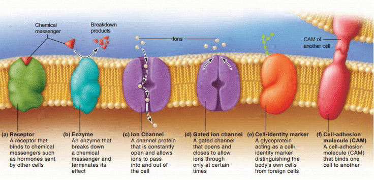
Fluid mosaic model: The membrane is a phospholipid bilayer containing mobile integral and peripheral proteins, cholesterol, glycolipids, and glycoproteins. Its fluidity permits transport, signalling, recognition, and membrane fusion; cholesterol buffers fluidity as temperature changes.
Membrane Lipids
Unsaturated fatty acid tails introduce bends that prevent tight packing, enhancing fluidity. Cholesterol acts as a temperature buffer in animal cells.
Membrane Proteins
Integral (Transmembrane) Proteins span the hydrophobic core. Functions include ion channels, carrier transporters, and signal receptors (e.g., GPCRs).
• Permeable (Unassisted): Small non-polar molecules ($\text{O}_2, \text{CO}_2, \text{N}_2$), hydrophobic steroid hormones.
• Weakly Permeable: Small polar uncharged molecules ($\text{H}_2\text{O}$, Glycerol, Urea).
• Impermeable (Requires Transporters): Large polar uncharged molecules (Glucose) and ALL charged ions ($\text{Na}^+, \text{K}^+, \text{Ca}^{2+}, \text{Cl}^-$).
1.3 Prokaryotes vs. Eukaryotes: Architectural Comparison
Living organisms divide into two fundamental cellular architectures: Prokaryotes and Eukaryotes. The defining evolutionary milestone of eukaryotes is internal cellular compartmentalization.
Figure 1.3A: Prokaryotic cell showing nucleoid and capsule.
Figure 1.3B: Compartmentalization vs simplicity.
Diagram: Prokaryotic Cell
Diagram: Eukaryotic Cell
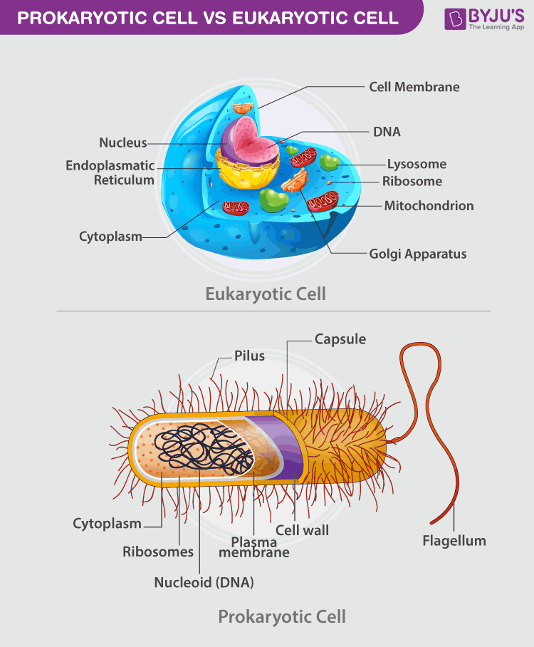
Prokaryotic cell: DNA lies in a nucleoid rather than a membrane-bound nucleus. Typical features include 70S ribosomes, a cell wall, plasmids, and sometimes a capsule or flagellum. Prokaryotes also lack membrane-bound organelles.
Eukaryotic cell: DNA is enclosed in a nucleus and cellular functions are compartmentalised in organelles such as mitochondria, ER, Golgi apparatus, and lysosomes. Cytosolic ribosomes are 80S.
| Feature | Prokaryotes (Bacteria & Archaea) | Eukaryotes (Animals, Plants, Fungi) |
|---|---|---|
| Nuclear Compartment | Absent. DNA lies in an unenclosed Nucleoid. | Present. True double-membrane Nucleus. |
| Genomic DNA Structure | Single circular chromosome; lacks histones (in Eubacteria); Plasmids present. | Multiple linear chromosomes wrapped around basic Histones. |
| Organelle Partitioning | Absent. No membrane-bound organelles. | Present. Extensive Endomembrane System and mitochondria. |
| Ribosomes | Smaller 70S Ribosomes (50S + 30S). | Larger cytosolic 80S Ribosomes (60S + 40S). |
| Cell Wall Polymer | Eubacteria: Peptidoglycan. | Plants: Cellulose. Fungi: Chitin. Animals: None. |
| Cell Division | Binary Fission (amitotic division). | Mitosis and Meiosis. |
1.4 Eukaryotic Organelles & Endomembrane Dynamics
Eukaryotic cells divide metabolic tasks among specialized membrane-enclosed organelles:
- Nucleus & Nucleolus: Enclosed by a double nuclear envelope. The dense Nucleolus synthesizes ribosomal RNA (rRNA).
- Rough ER (RER): Studded with 80S ribosomes. Synthesizes proteins destined for secretion or membranes.
- Smooth ER (SER): Lacks ribosomes. Responsible for lipid synthesis and drug detoxification (e.g., in liver cells).
- Golgi Apparatus: Modifies, sorts, and packages proteins into vesicles (the "post office").
- Lysosomes: Acidic vesicles filled with hydrolytic enzymes for intracellular digestion and autophagy.
- Mitochondria: Double-membrane powerhouses. The site of cellular respiration to generate ATP. Contains its own DNA.
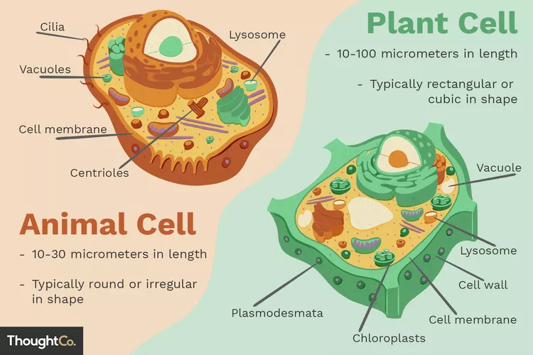
Special Structures in Plant Cells
Plant cells are eukaryotic but possess structures absent in animal cells:
Diagram: Animal Cell vs. Plant Cell
- Cell Wall: Rigid external layer made of cellulose. Prevents osmotic lysis.
- Chloroplasts: Site of photosynthesis. Contain chlorophyll.
- Large Central Vacuole: Stores water/nutrients and maintains turgor pressure.
Part 2: The Code of Life - DNA, RNA, and Protein Synthesis
2.1 Nucleotide Chemistry & Nucleic Acid Architecture
The DNA molecule is the master blueprint. It is a polymer made of repeating units called nucleotides. Each nucleotide contains three constituents:

Figure 2.1A: DNA Molecular Structure overview.
- Pentose Sugar: $2'$-deoxyribose in DNA vs ribose in RNA.
- Phosphate Group: Forms the phosphodiester sugar-phosphate backbone.
- Nitrogenous Base: The "letters" of the genetic code.
Diagram: Structure of a DNA Nucleotide
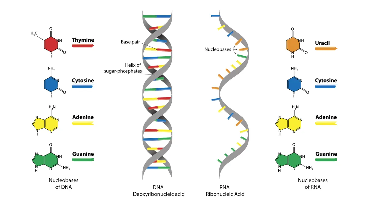
DNA nucleotide: Each nucleotide contains phosphate, deoxyribose, and one nitrogenous base. Adjacent nucleotides form phosphodiester bonds, while complementary bases pair through hydrogen bonds: A–T has two and G–C has three.
Purine Bases (Double Ring)
Includes Adenine (A) and Guanine (G). Present in both DNA and RNA.
Pyrimidine Bases (Single Ring)
Includes Cytosine (C), Thymine (T) [DNA only], and Uracil (U) [RNA only].
2.2 DNA Double Helix & Chargaff's Rules
DNA forms an anti-parallel double helix held together by complementary hydrogen bonding.
Figure 2.2: Watson-Crick anti-parallel double helix model.
$$\text{Adenine (A)} = \text{Thymine (T)} \quad (\text{linked by 2 Hydrogen Bonds})$$
$$\text{Guanine (G)} = \text{Cytosine (C)} \quad (\text{linked by 3 Hydrogen Bonds})$$
$$\text{Total Purines } (A+G) = \text{Total Pyrimidines } (T+C) = 50\%$$
2.3 Chromatin Hierarchy & Genomic Packaging
To fit inside the nucleus, linear DNA undergoes systematic folding into **chromatin**:
Diagram: DNA Packaging into a Chromosome
- Nucleosome: DNA wrapped around a basic histone octamer.
- Euchromatin: Loosely packed, transcriptionally active.
- Heterochromatin: Highly condensed, transcriptionally silent.
2.4 DNA Replication Mechanism
Occurs during the S Phase via a semiconservative mechanism.
Diagram: DNA Replication Fork
1. DNA Helicase: Unwinds the double helix.
2. Primase: Synthesizes short RNA primers.
3. DNA Polymerase III: Elongates new strands ($5' \rightarrow 3'$).
4. DNA Ligase: Seals nicks between Okazaki Fragments on the lagging strand.
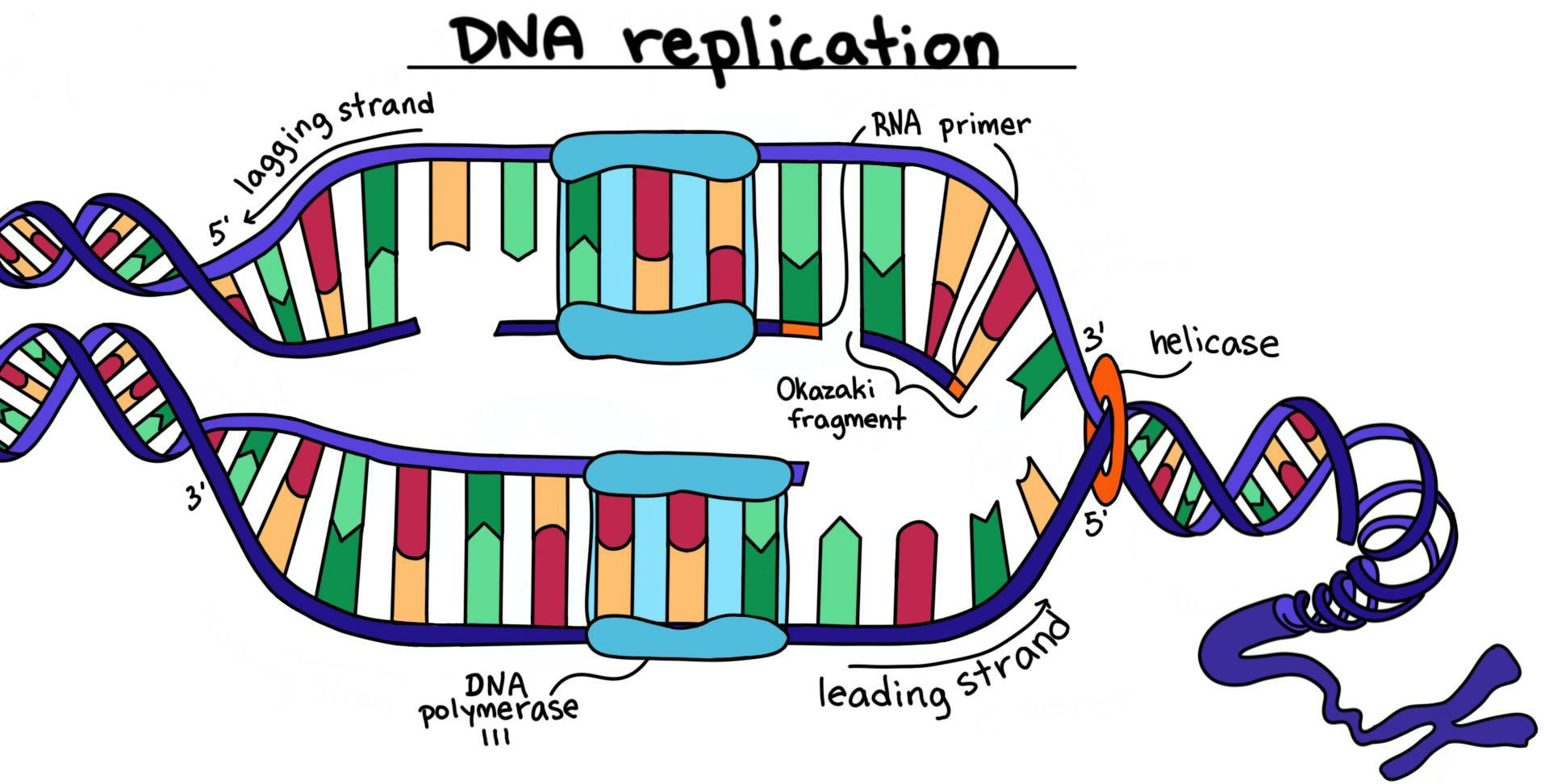
Replication fork: DNA polymerase synthesises only in the 5′→3′ direction. The leading strand is continuous; the lagging strand forms Okazaki fragments that are later joined by DNA ligase. Helicase separates the parental strands and primase supplies RNA primers.
2.5 The Central Dogma: Transcription & Translation
The Central Dogma describes the flow: $\text{DNA} \xrightarrow{\text{Transcription}} \text{RNA} \xrightarrow{\text{Translation}} \text{Protein}$.

Diagram: Transcription and Translation
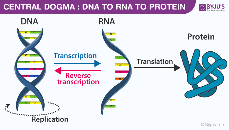
Transcription: RNA polymerase reads the DNA template strand and synthesises complementary RNA. In eukaryotes, pre-mRNA is processed by 5′ capping, polyadenylation, and splicing before leaving the nucleus.
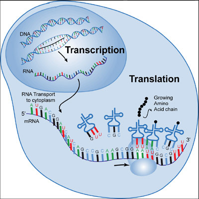
Translation: The ribosome reads mRNA codons and tRNAs deliver the corresponding amino acids. Peptide bonds form as tRNAs move through the A, P, and E sites; AUG is the usual start codon.
Part 3: The Energy Engine - Metabolism
3.1 Catabolism, Anabolism, and ATP
Metabolism manages material and energy resources via two processes:

- Catabolism ($\Delta G < 0$): Exergonic breakdown (e.g., Cellular respiration) releasing energy.
- Anabolism ($\Delta G > 0$): Endergonic synthesis (e.g., Photosynthesis) requiring energy.
Diagram: The ATP-ADP Energy Cycle
$$\text{ATP} + \text{H}_2\text{O} \rightarrow \text{ADP} + \text{P}_i + \text{Energy} \quad (\Delta G^\circ = -30.5\text{ kJ/mol})$$
3.2 Aerobic Respiration vs. Photosynthesis Integration
These two form Earth's fundamental bioenergetic feedback loop.
Diagram: The Cycle of Photosynthesis and Respiration
Photosynthesis: $6\text{CO}_2 + 6\text{H}_2\text{O} + \text{Light Energy} \rightarrow \text{C}_6\text{H}_{12}\text{O}_6 + 6\text{O}_2$
Four Stages of Aerobic Respiration:
- Glycolysis (Cytosol): Glucose → 2 Pyruvate. Net: 2 ATP, 2 NADH.
- Pyruvate Oxidation (Matrix): → Acetyl-CoA + $CO_2$ + NADH.
- Krebs Cycle (Matrix): Yields 2 ATP, 6 NADH, 2 $FADH_2$, $4 CO_2$.
- Oxidative Phosphorylation (Inner Membrane): ETC powers ATP Synthase. Terminal acceptor is $O_2$. Yields $\approx 28\text{ ATP}$.
Part 4: The Cell Cycle & Cellular Division

4.1 Eukaryotic Cell Cycle & Control Checkpoints
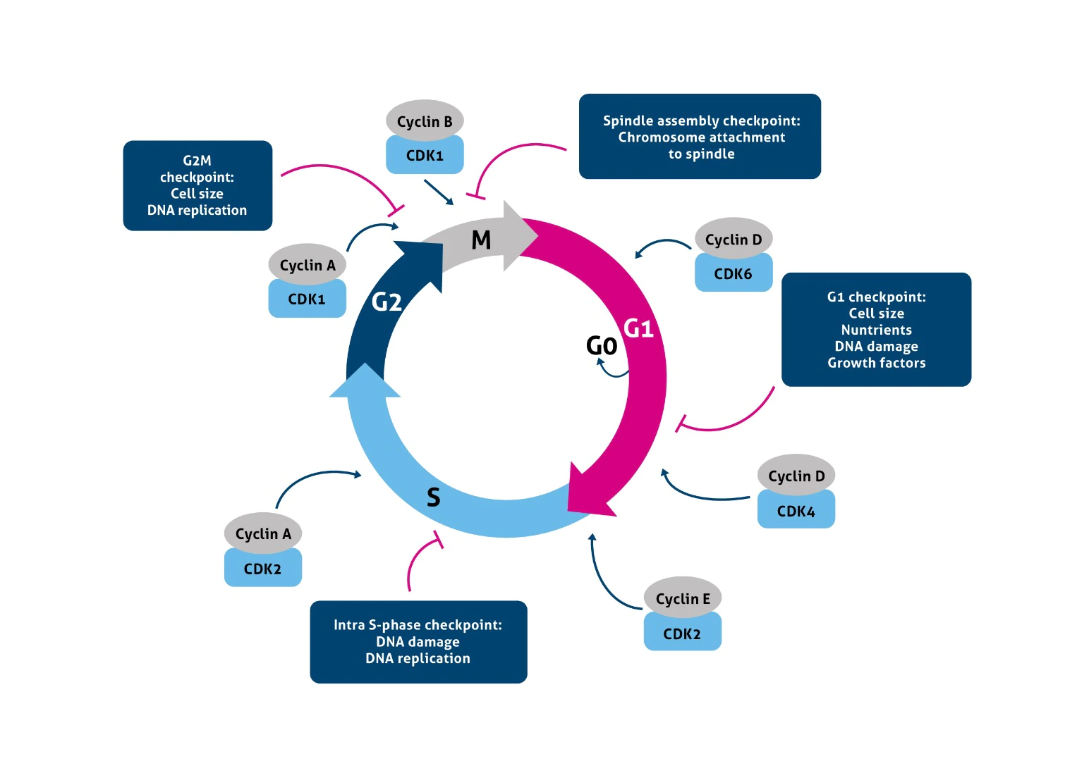
Diagram: The Eukaryotic Cell Cycle
- Interphase (90%): Comprises $G_1$ (growth), $S$ (DNA replication), and $G_2$ (division prep).
- M Phase: Mitosis (nuclear division) and Cytokinesis.
Cell Cycle Control System (Cyclins & CDKs):
Progression is strictly regulated by Cyclin-Dependent Kinases (CDKs) activated by regulatory Cyclin proteins. Checkpoints ($G_1$, $G_2$, $M$) ensure genomic integrity.
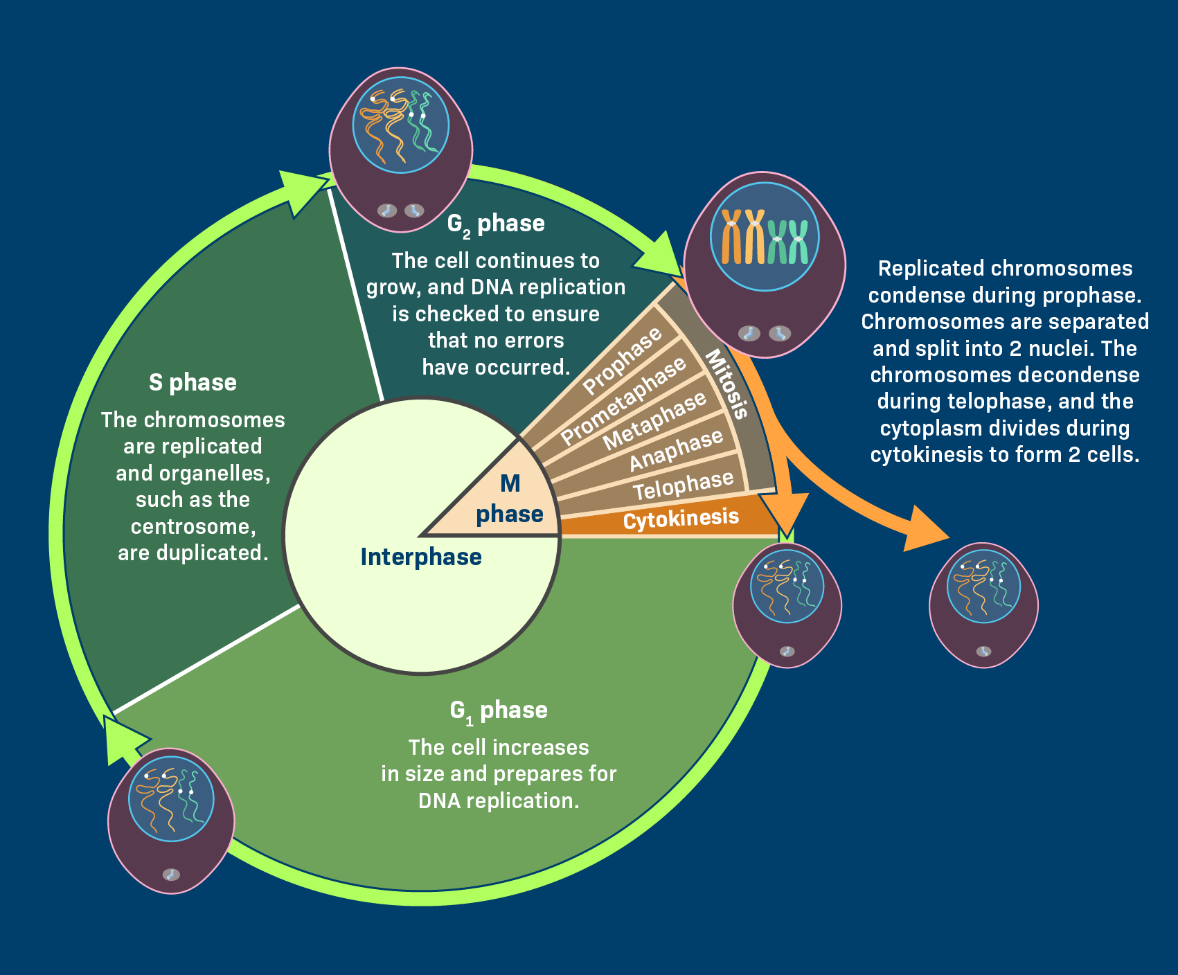
Cell cycle: Interphase consists of G₁, S, and G₂, followed by mitosis and cytokinesis. Checkpoints at G₁/S, G₂/M, and metaphase prevent damaged DNA or incorrectly attached chromosomes from being passed on.
Mitosis: Chromosomes condense in prophase, align at the metaphase plate, sister chromatids separate in anaphase, and nuclei reform in telophase. Cytokinesis then divides the cytoplasm.
4.2 Mitosis vs. Meiosis Mechanics
Figure 4.1: Mitosis vs Meiosis direct comparison.
| Parameter | Mitosis (Equational) | Meiosis (Reductional & Equational) |
|---|---|---|
| Primary Purpose | Somatic growth, tissue repair. | Gametogenesis (Sperm and Ovum production). |
| Rounds of Division | 1 Division. | 2 Divisions (Meiosis I and II). |
| Daughter Cell Yield | 2 Diploid ($2n$) genetically identical. | 4 Haploid ($n$) genetically unique gametes. |
| Variation generation | Absent. | Crossing Over and Independent Assortment. |
Part 5: Practice Questions and Review
Test your understanding of Lesson 0 content. Select your answers and click Submit Answers.