Lesson 6: Cell Structure & Membrane Transport
Build the cell as a connected system: architecture → information flow → protein trafficking → energy → mechanics → junctions → membrane transport → organelle atlas & cell diversity → viruses. This revised version is intentionally denser and more colorful, adding deep explanations of centrosomes, vacuoles, proteasomes, endosymbiotic evidence, cell fractionation, organelle sizes, organism-by-organism organelle distribution, and high-yield specialized-cell exceptions that are often tested.
Learning Objectives
The primary cellular divide is between prokaryotes (Bacteria and Archaea) and eukaryotes. The difference is not simply “small versus large”: eukaryotes use membrane-bound compartments to separate incompatible reactions and create specialized microenvironments.
1.1 Prokaryotic Cell: Compact but Highly Organized

Cellular architectures: Use this image to compare nucleoid vs nucleus, 70S vs 80S ribosomes, compartmentalization, cytoskeletal complexity and the bacterial origins of mitochondria/chloroplasts.
Gram-Positive vs Gram-Negative: Read the Envelope from Outside → Inside
1.2 Eukaryotic Cells & Endosymbiotic Theory
Eukaryotic cells contain a true nucleus, an extensive endomembrane system and multiple specialized organelles. Endosymbiotic theory proposes that an ancestral host cell engulfed bacteria that were not digested and instead became stable endosymbionts. Over evolutionary time the partnership became obligatory: many genes moved to the host nucleus, but mitochondria and chloroplasts still retain enough bacterial features to reveal their origin.

Animal cell: Note the nucleus, ER, Golgi, mitochondria, lysosomal/endosomal compartments and cytoskeleton.

Plant cell: The eukaryotic core is supplemented by a cellulose wall, chloroplasts, plasmodesmata and a large central vacuole.
🧫 Endosymbiosis — the sequence, not a text-heavy picture
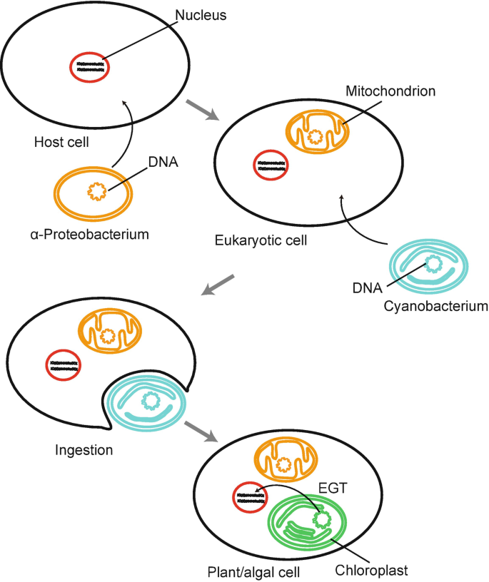
Endosymbiotic theory: The transition from free-living bacterium to permanent organelle explains why mitochondria and chloroplasts carry a double membrane, bacterial-type ribosomes, circular DNA and a division mode reminiscent of binary fission.
| Evidence | What we observe | Why it supports endosymbiosis | High-yield note |
|---|---|---|---|
| Double membrane | Mitochondria and chloroplasts have outer and inner membranes. | The inner membrane resembles the ancestral bacterial membrane; the outer membrane reflects engulfment by the host. | Chloroplast thylakoids are an additional internal membrane system, not the same thing as the envelope. |
| Circular DNA | Both organelles contain small circular genomes. | Circular genomes are characteristic of bacteria, unlike the linear chromosomes of the nucleus. | Organelle DNA is limited; many ancestral genes have moved to the nucleus. |
| 70S ribosomes | Mitochondria/chloroplasts translate some proteins using bacterial-sized ribosomes. | These ribosomes are closer to bacterial ribosomes than to the 80S cytosolic ribosomes of eukaryotes. | This is why some antibiotics that affect bacteria can also affect mitochondrial translation. |
| Binary-fission-like division | Organelles enlarge and divide rather than being synthesized de novo. | Division resembles bacterial binary fission. | They still depend on host genes, so they are semiautonomous only. |
| Gene/protein homology | Sequence analysis links mitochondria to α-proteobacteria and chloroplasts to cyanobacteria. | Molecular phylogeny independently confirms the structural evidence. | Phylogeny is the strongest modern support because it compares many genes, not a single feature. |
| Feature | Prokaryotes | Eukaryotes | IMAT anchor |
|---|---|---|---|
| Nucleus | No nuclear envelope; nucleoid | Double-membrane nucleus | Nuclear envelope = eukaryotic compartmentalization |
| Genome | Usually circular main chromosome; plasmids may occur | Multiple linear chromosomes with histones | Do not confuse bacterial plasmids with the main chromosome |
| Ribosomes | 70S | 80S cytosolic; 70S in mitochondria/chloroplasts | Endosymbiotic evidence |
| Organelles | No classic membrane-bound organelles | ER, Golgi, mitochondria, lysosomes, etc. | Compartmentalization enables specialization |
| Division | Binary fission | Mitosis / meiosis | FtsZ is a tubulin homolog |
Cell architecture
Apply this section immediately with official IMAT past-paper questions. Select an answer to reveal the full worked explanation.
Which of the following statements accurately describes prokaryotic DNA?
The decisive statement is Prokaryotic DNA consists of circular chromosomes located within the cytoplasm.
This question tests Cell architecture. Apply the definitions, structural relationships, and cause-and-effect rules in the section immediately above; the remaining choices conflict with at least one of those conditions.
Test every condition in the stem independently. Reject an option as soon as one statement contradicts the biological rules established in this lesson.
Which of the following are features of all prokaryotes?
1 They have a cell membrane.
2 They have a nucleus.
3 They have ribosomes.
4 They have mitochondria.
Which of the following statements about all prokaryotes are correct?
1 They contain DNA.
2 They are single-celled.
3 They have 70S ribosomes.
4 They have a cell wall.
2. Single-celled: True. There are absolutely no multicellular prokaryotic organisms. They may form biofilms or colonies, but each cell is an independent organism.
3. 70S Ribosomes: True. This is the defining ribosome size and density for prokaryotes.
Which characteristic is found in all members of the Kingdom Monera?
C: Many bacteria lack flagella or pili and are completely non-motile (e.g., many cocci forms).
D: As mentioned in previous explanations, bacteria of the genus Mycoplasma lack a cell wall altogether, making peptidoglycan non-universal.
E: While *most* bacteria have a single, circular chromosome, there are rare but well-documented exceptions (like Borrelia burgdorferi, the causative agent of Lyme disease, and Streptomyces) which possess linear chromosomes. "Absence of a nucleus" is the only universally safe answer.
Four students, 1, 2, 3 and 4, recorded different structures that they thought were found in a healthy animal kidney cell and a typical bacterial cell. Which students gave totally correct answers?
| student | animal cell | bacterial cell |
|---|---|---|
| 1 | DNA found in a nucleus | 70S ribosomes present |
| 2 | cell wall present | mitochondria present |
| 3 | 70S ribosomes present | DNA found in a nucleus |
| 4 | circular DNA | plasmids present |
The decisive statement is students 1 and 4 only
This question tests Cell architecture. Apply the definitions, structural relationships, and cause-and-effect rules in the section immediately above; the remaining choices conflict with at least one of those conditions.
Test every condition in the stem independently. Reject an option as soon as one statement contradicts the biological rules established in this lesson.
Two disease-free cells, P and Q, were studied. Which option correctly identifies cell P as a typical plant cell and cell Q as a typical prokaryote?
| Cell P | Cell Q | |
|---|---|---|
| Row 1 | Gene for RuBisCo is present | Susceptible to penicillin |
| Row 2 | Plasmids present | Centrioles found as a pair |
| Row 3 | Outermost layer selectively permeable | SER present |
| Row 4 | Glycogen can be present | Cell wall is present |
| Row 5 | Organelle with grana present | Nucleolus may be present |
The decisive statement is Row 1
This question tests Cell architecture. Apply the definitions, structural relationships, and cause-and-effect rules in the section immediately above; the remaining choices conflict with at least one of those conditions.
Test every condition in the stem independently. Reject an option as soon as one statement contradicts the biological rules established in this lesson.
Which of the following healthy cells contain circular DNA?
1 Escherichia coli
2 Saccharomyces cerevisiae (yeast)
3 human liver cell
The decisive statement is 1, 2 and 3
This question tests Cell architecture. Apply the definitions, structural relationships, and cause-and-effect rules in the section immediately above; the remaining choices conflict with at least one of those conditions.
Test every condition in the stem independently. Reject an option as soon as one statement contradicts the biological rules established in this lesson.
The presence of intercellular compartmentalisation is a characteristic of which organisms?
The decisive statement is Of eukaryotes
This question tests Cell architecture. Apply the definitions, structural relationships, and cause-and-effect rules in the section immediately above; the remaining choices conflict with at least one of those conditions.
Test every condition in the stem independently. Reject an option as soon as one statement contradicts the biological rules established in this lesson.
Which of the following statements about a neurone in a healthy human is/are correct?
1 Transport across its cell membrane can occur by exocytosis.
2 It contains the gene that codes for insulin.
3 It contains circular DNA.
2. Insulin Gene: All somatic (diploid body) cells in a human contain the exact same complete genome. Therefore, a brain cell absolutely contains the gene for insulin. It is simply switched off (methylated/repressed) so the neuron doesn't actually produce insulin. (True)
3. Circular DNA: Neurons are highly active cells packed with mitochondria to produce ATP. According to endosymbiotic theory, all mitochondria contain their own loop of circular DNA (mtDNA). Therefore, the neuron contains circular DNA inside its mitochondria. (True)
2.1 Nuclear Architecture
🧠 Think of the nucleus as a controlled information environment
The nucleus is not simply a container for DNA. Its envelope physically separates transcription and RNA processing from cytoplasmic translation. That separation allows eukaryotic cells to splice pre-mRNA, control which RNAs leave the nucleus, and regulate access to DNA with much greater precision than a prokaryotic cell.
The inner nuclear membrane is mechanically supported by lamins, while chromatin is tethered to specific regions of the nuclear periphery. Nuclear pore complexes then act as selective gates. Small solutes can diffuse, but large proteins and RNAs require transport receptors and directional Ran-GTP chemistry.
The nucleus separates DNA replication, transcription and RNA processing from cytoplasmic translation. The outer nuclear membrane is continuous with rough ER, while the inner surface is supported by the nuclear lamina, a network of lamin intermediate filaments.

Nucleus and traffic: Focus on the double membrane, NPC, lamina and directional Ran-GTP system rather than treating the nucleus as a static DNA container.
Nuclear Import: NLS → Importin → NPC → Ran-GTP
2.2 Nucleolus & Ribosome Biogenesis
The nucleolus is a non-membranous nuclear body where rRNA is transcribed/processed and ribosomal subunits are assembled. Eukaryotic cytosolic ribosomes are 80S = 60S + 40S. Their functional sites are A (aminoacyl), P (peptidyl), and E (exit).
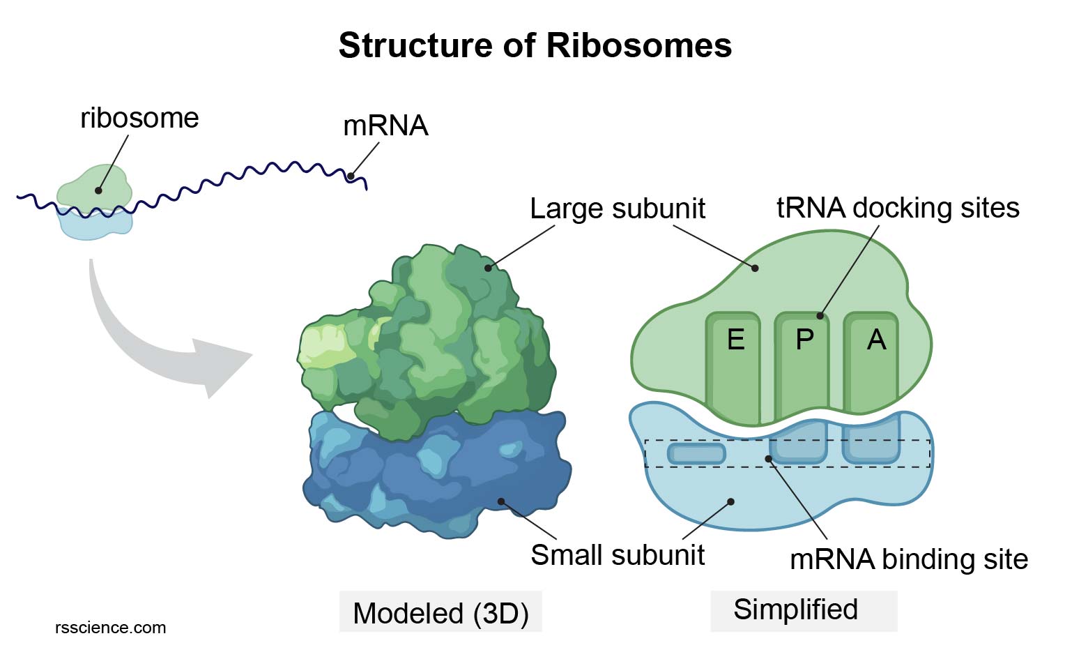
Ribosome architecture: Large and small subunits create the mRNA/tRNA interface used during translation.
Ribosome Logic: A → P → E
| Structure | Composition / location | Main role | High-yield distinction |
|---|---|---|---|
| Nuclear envelope | Inner + outer membranes | Separates nuclear processes | Outer membrane is continuous with ER |
| Lamina | Lamins (intermediate filaments) | Mechanical support, chromatin organization | Not microtubules or actin |
| NPC | Nucleoporins | Selective nucleo-cytoplasmic traffic | Large cargo uses transport receptors |
| Nucleolus | Non-membranous | rRNA + ribosome assembly | Dense but not membrane-bound |
The endomembrane system is best understood as a logistics network. Secretory and membrane proteins are synthesized into the ER, checked for quality, moved to Golgi, modified, sorted and then delivered to the plasma membrane, secretory vesicles, endosomes or lysosomes.

Secretory pathway: Follow cargo from RER to cis-Golgi, medial/trans stacks and the TGN. Destination depends on sorting signals, not on random vesicle movement.
Secretory Protein Journey: From Ribosome to Destination
3.1 Rough ER vs Smooth ER
🏭 The ER is a continuous membrane system with different functional zones
The rough and smooth ER are connected regions of the same organelle. A ribosome becomes “ER-bound” only when the protein being synthesized contains an ER signal sequence. Signal-recognition particle (SRP) pauses translation, docks the ribosome to the SRP receptor, and positions the nascent chain at the SEC61 translocon. Translation then resumes while the polypeptide enters the ER lumen or membrane.
Inside the rough ER, chaperones such as BiP assist folding, disulfide bonds form, and N-linked glycans are added to asparagine residues. The calnexin/calreticulin quality-control cycle repeatedly tests glycoprotein folding. Proteins that cannot reach a stable native state are retrotranslocated into the cytosol, ubiquitinated and destroyed by the proteasome through ERAD. Persistent accumulation of unfolded proteins activates PERK, IRE1 and ATF6 branches of the unfolded-protein response.
The smooth ER has few bound ribosomes and therefore emphasizes lipid and steroid synthesis, drug detoxification through cytochrome P450 enzymes, and Ca²⁺ handling. In skeletal and cardiac muscle it becomes the sarcoplasmic reticulum, where rapid Ca²⁺ release and reuptake directly control contraction.
| Feature | Rough ER | Smooth ER |
|---|---|---|
| Surface | Ribosomes attached | No ribosomes |
| Main products | Secreted, lysosomal and membrane proteins | Lipids, phospholipids, steroid precursors |
| Special functions | Folding, disulfides, N-linked glycosylation, quality control | Drug detoxification, Ca²⁺ storage, lipid metabolism |
| Clinical/IMAT anchor | Misfolded proteins → ERAD / UPR | Sarcoplasmic reticulum = specialized SER |

ER quality control: Chaperones help proteins fold; irreversibly misfolded proteins can be retrotranslocated, ubiquitinated and degraded by the proteasome. Persistent stress activates the UPR.
3.2 Golgi Apparatus: Modification & Sorting
The Golgi is polarized: cis receives ER cargo, medial cisternae modify it, and the trans-Golgi network performs final sorting. N-linked glycans are remodeled; O-linked glycosylation occurs predominantly in the Golgi.

Golgi: Think cis → medial → trans. Cisternae mature while resident enzymes are recycled backward.
3.3 Vesicle Coats, SNAREs, Endosomes & Lysosomes
| Coat / signal | Direction | Core function | Memory rule |
|---|---|---|---|
| COPII | ER → Golgi | Anterograde secretory traffic | “II = out of ER” |
| COPI | Golgi → ER / intra-Golgi | Retrograde retrieval | Returns machinery and ER residents |
| Clathrin | TGN → endosome; PM → endosome | Lysosomal sorting and receptor-mediated endocytosis | Selective cargo concentration |
| SNAREs | Vesicle + target membrane | Specific membrane fusion | v-SNARE pairs with t-SNARE |
| M6P | Golgi → endosome/lysosome | Targets acid hydrolases | Lysosomal “zip code” |
Endomembrane system
Apply this section immediately with official IMAT past-paper questions. Select an answer to reveal the full worked explanation.
Which of the following organelles would be expected to be found in both photosynthetic plant cells and non-photosynthetic animal cells?
1 Rough endoplasmic reticulum (RER)
2 Ribosomes
3 Mitochondria
4 Cell wall
The decisive statement is 1, 2 and 3 only
This question tests Endomembrane system. Apply the definitions, structural relationships, and cause-and-effect rules in the section immediately above; the remaining choices conflict with at least one of those conditions.
Test every condition in the stem independently. Reject an option as soon as one statement contradicts the biological rules established in this lesson.
Which of the following organelles are bounded by a single membrane?
1 Lysosome
2 Mitochondrion
3 Golgi apparatus
4 Ribosome
The decisive statement is 1 and 3 only
This question tests Endomembrane system. Apply the definitions, structural relationships, and cause-and-effect rules in the section immediately above; the remaining choices conflict with at least one of those conditions.
Test every condition in the stem independently. Reject an option as soon as one statement contradicts the biological rules established in this lesson.
Which of the following organelles is NOT directly involved in the synthesis or processing of proteins intended for secretion outside the cell?
The decisive statement is Smooth endoplasmic reticulum (SER)
This question tests Endomembrane system. Apply the definitions, structural relationships, and cause-and-effect rules in the section immediately above; the remaining choices conflict with at least one of those conditions.
Test every condition in the stem independently. Reject an option as soon as one statement contradicts the biological rules established in this lesson.
4.1 Mitochondria
⚡ Structure follows energy function
Mitochondria are double-membrane organelles whose internal architecture creates two chemically different spaces: the matrix and the intermembrane space. The inner membrane is extremely protein-rich and almost impermeable to ions, which is essential because the electron transport chain uses it to maintain a proton electrochemical gradient.
Cristae dramatically expand inner-membrane surface area. Cells with high and continuous ATP demand—such as cardiomyocytes—therefore contain many mitochondria with dense cristae. The matrix contains pyruvate dehydrogenase, most TCA-cycle enzymes, β-oxidation machinery, mitochondrial DNA and mitochondrial ribosomes. Mitochondria also regulate apoptosis, Ca²⁺ buffering and metabolic signaling, so “powerhouse” is only part of the story.
Mitochondria are double-membrane organelles that couple fuel oxidation to ATP production. Their compartments are functionally different, so IMAT questions often ask where a reaction occurs.

Metabolic hubs: Match mitochondrial cristae with oxidative phosphorylation and matrix with Krebs-cycle/fatty-acid metabolism; contrast this with peroxisomal oxidative reactions.
⚡ Mitochondrial Compartment Map — labels kept outside the organelle
4.2 Chloroplasts
Chloroplasts contain an outer and inner envelope plus an internal thylakoid membrane. Light-dependent reactions occur in the thylakoid membrane; the Calvin cycle occurs in the stroma.

Chloroplast: Distinguish thylakoids/grana from stroma. Chloroplast DNA and 70S ribosomes provide additional endosymbiotic evidence.
4.3 Peroxisomes
Peroxisomes are single-membrane oxidative organelles. In addition to β-oxidation of very-long-chain fatty acids, they carry out α-oxidation of branched fatty acids (for example phytanic acid), participate in plasmalogen synthesis important for myelin, and contribute to bile-acid and reactive-oxygen metabolism. Oxidases generate hydrogen peroxide during oxidation, and catalase rapidly destroys excess H₂O₂ before it damages the cell.
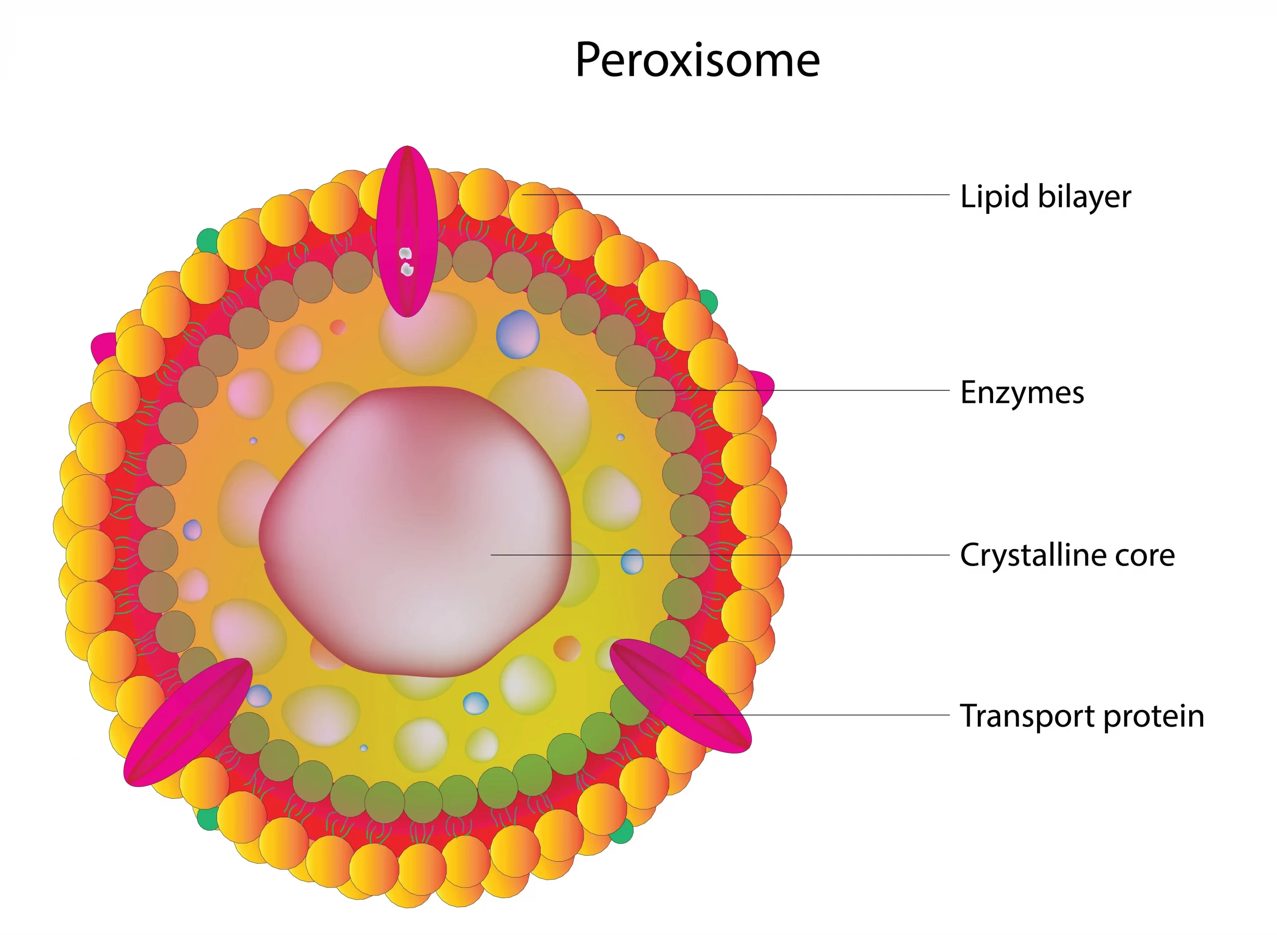
Peroxisome: Peroxisomes do not make ATP by oxidative phosphorylation and they do not contain their own DNA. Their identity is oxidative metabolism, detoxification and peroxide handling. Zellweger-spectrum disorders and X-linked adrenoleukodystrophy are classic clinical associations.
Peroxisomal Detox Logic
Energy organelles
Apply this section immediately with official IMAT past-paper questions. Select an answer to reveal the full worked explanation.
Which process occurs within mitochondria?
C: Photosynthesis occurs in chloroplasts.
E: Microbodies (such as peroxisomes) generally form by budding off from the Endoplasmic Reticulum, not within mitochondria.
The cytoskeleton is not a fixed skeleton. Actin and microtubules constantly assemble/disassemble, allowing a cell to change shape, divide, transport cargo and build specialized structures.
| Filament | Subunit | Polarity | Characteristic dynamics | Major functions |
|---|---|---|---|---|
| Actin / microfilament | G-actin → F-actin | Polar | ATP-dependent treadmilling | Cortex, motility, microvilli, cytokinesis, myosin contraction |
| Microtubule | α/β-tubulin dimers | Polar | GTP-cap dynamic instability | Organelle transport, spindle, cilia/flagella |
| Intermediate filament | Keratin, vimentin, desmin, lamins, etc. | Non-polar | Relatively stable | Tensile strength, desmosomes, nuclear lamina |

Cytoskeleton: Use the image to connect actin with myosin, microtubules with kinesin/dynein, and intermediate filaments with mechanical resilience.
Motor Directionality & Cytoskeletal Tracks

Motor proteins: Microtubule polarity allows kinesin and dynein to move cargo in opposite directions along the same track.
5.1 Treadmilling vs Dynamic Instability
🛤️ The cytoskeleton is a moving transport network, not a static skeleton
Actin filaments and microtubules are polar polymers, meaning their two ends are chemically different. This polarity gives the cell direction. Myosins read actin polarity, whereas kinesins and dyneins read microtubule polarity. As a result, cells can route vesicles, organelles and chromosomes to specific destinations instead of relying on random diffusion.
Actin often shows treadmilling: subunits can be added preferentially at the plus end while older subunits leave the minus end. Microtubules instead show dynamic instability, switching between growth and rapid shrinkage according to the state of the GTP cap. These dynamic behaviors are crucial for cell migration, mitotic spindle searching and reorganization of cell shape.
5.2 Cilia & Flagella
Motile eukaryotic cilia and flagella contain a 9+2 axoneme: nine outer microtubule doublets around two central singlets. Axonemal dynein generates sliding forces; nexin links and structural constraints convert sliding into bending.
9+2 Axoneme
6.1 Animal Cell Junctions
🧱 A tissue needs seals, rivets, belts and communication channels
Different junctions solve different mechanical or physiological problems. Tight junctions control leakage between cells and help preserve apical-basal polarity. Adherens junctions connect cadherins to cortical actin and allow a sheet of cells to change shape. Desmosomes connect intermediate-filament networks and therefore resist tearing. Gap junctions do the opposite of sealing: they create aqueous channels that directly couple neighboring cytoplasms for ions and small signaling molecules.
Do not memorize the names without the cytoskeletal link. Tight = barrier, adherens = actin belt, desmosome = intermediate-filament rivet, gap = channel.

Cell junctions: The core distinction is functional: tight = seal, adherens = actin belt, desmosome = IF rivet, gap = communication channel.

Dedicated junction summary: Compare tight junctions, adherens junctions, desmosomes and gap junctions by the protein complex involved, the cytoskeletal element attached, and whether the purpose is sealing, adhesion or communication.
Junction Ladder: From Apical Seal to Basal Attachment
| Junction | Main proteins | Cytoskeletal link | Function |
|---|---|---|---|
| Tight | Claudins, occludins | Actin-associated scaffold | Seal paracellular pathway; maintain polarity |
| Adherens | Cadherins + catenins | Actin | Adhesion belt; tissue shape |
| Desmosome | Desmoglein, desmocollin | Intermediate filaments | Strong spot adhesion |
| Gap | Connexins → connexons | No major anchoring role | Pass ions/small molecules between cells |
6.2 ECM & Integrins

ECM: Collagen gives tensile strength; proteoglycans hold water; fibronectin links ECM to integrins. Integrins connect ECM to cytoskeleton and transmit mechanical/chemical signals.
6.3 Plant Cell Structures
7.1 Fluid Mosaic Model & Selective Permeability
🚪 Membrane transport starts with chemistry
The phospholipid bilayer has a hydrophobic interior. Small non-polar molecules dissolve in that interior and cross readily, whereas ions and large polar molecules face a strong energetic barrier. Membrane proteins solve that problem by providing selective channels, carriers or pumps.
A concentration gradient is only part of the story for ions. Because ions carry charge, movement is governed by an electrochemical gradient: the combined effect of concentration difference and membrane voltage. A transporter can therefore move a solute downhill chemically but uphill electrically, or vice versa. This is why secondary active transport depends on stored gradient energy rather than direct ATP hydrolysis by the cotransporter itself.
The membrane is a dynamic phospholipid bilayer containing proteins, cholesterol and carbohydrates. Hydrophobicity, size and charge determine whether a molecule can cross the lipid bilayer directly.

Membrane and transport: Use this image as a reference while separating passive transport, active transport and bulk vesicular transport into distinct mechanisms.
| 🧪 Molecule class | 🚪 Bilayer crossing | 🛣️ Typical route | 🔎 Example |
|---|---|---|---|
| Small non-polar | Easy | Simple diffusion directly through the hydrophobic core | O₂, CO₂, steroid molecules |
| Small polar | Limited | Often requires channels; water commonly uses aquaporins | H₂O |
| Ions | Essentially blocked | Ion channels, carriers, pumps, symporters or antiporters | Na⁺, K⁺, Ca²⁺, Cl⁻ |
| Large polar | Blocked | Specific carriers or vesicular transport depending on size | Glucose, peptides, proteins |

7.2 Transport Decision Tree

Transport classes: First ask whether the solute moves down or against its electrochemical gradient; then ask whether a membrane protein or direct energy input is required.
Choose the Mechanism by Asking Three Questions
7.3 Passive Transport & Osmosis
Simple diffusion crosses the bilayer directly. Facilitated diffusion still moves down the gradient but requires channels or carriers. Osmosis is net water movement across a selectively permeable membrane.

Animal cells: Hypotonic → water enters → swelling/lysis; isotonic → no net volume change; hypertonic → water leaves → crenation.

Plant cells: Hypotonic water entry creates turgor because the wall resists expansion; hypertonic solution causes plasmolysis.
Tonicity: Predict Water, Then Predict Cell Shape
7.4 Primary & Secondary Active Transport

Na⁺/K⁺ ATPase: One ATP moves 3 Na⁺ out and 2 K⁺ in. The pump is electrogenic and creates gradients used by many secondary transporters.

Pump mechanism: Phosphorylation and dephosphorylation change transporter conformation so ions bind on one side and are released on the other.
Primary Pump Creates the Gradient; Secondary Transport Spends It
7.5 Bulk Transport

Bulk transport: Exocytosis adds vesicle membrane to the plasma membrane; endocytosis removes membrane while bringing cargo inward.

Receptor-mediated endocytosis: Ligand-bound receptors cluster in clathrin-coated pits; dynamin pinches the vesicle free. Specificity comes from receptor–cargo recognition.
| Mechanism | Gradient / energy | Protein required? | Typical cargo |
|---|---|---|---|
| Simple diffusion | Down gradient; no ATP | No | O₂, CO₂, lipid-soluble molecules |
| Facilitated diffusion | Down electrochemical gradient | Channel or carrier | Ions, glucose |
| Primary active | Against gradient; ATP directly | Pump | Na⁺, K⁺, Ca²⁺, H⁺ |
| Secondary active | One solute downhill drives another uphill | Symporter/antiporter | Na⁺-glucose, Na⁺-Ca²⁺ exchange |
| Vesicular | Energy-dependent membrane remodeling | Coats/fusion machinery | Macromolecules, particles, secretory cargo |
Membrane transport
Apply this section immediately with official IMAT past-paper questions. Select an answer to reveal the full worked explanation.
What are carrier proteins?
The decisive statement is They are the proteins that transfer molecules and ions across the plasma membrane.
This question tests Membrane transport. Apply the definitions, structural relationships, and cause-and-effect rules in the section immediately above; the remaining choices conflict with at least one of those conditions.
Test every condition in the stem independently. Reject an option as soon as one statement contradicts the biological rules established in this lesson.
The graph shows the rate of uptake of a substance by a cell against the external concentration of the substance. Which method(s) of transport could be represented by this graph?
1 Simple diffusion
2 Facilitated diffusion
3 Active transport
The decisive statement is 2 and 3 only
This question tests Membrane transport. Apply the definitions, structural relationships, and cause-and-effect rules in the section immediately above; the remaining choices conflict with at least one of those conditions.
Test every condition in the stem independently. Reject an option as soon as one statement contradicts the biological rules established in this lesson.
Active transport, facilitated diffusion and simple diffusion share the feature of:
The decisive statement is transport across the cell surface membrane.
This question tests Membrane transport. Apply the definitions, structural relationships, and cause-and-effect rules in the section immediately above; the remaining choices conflict with at least one of those conditions.
Test every condition in the stem independently. Reject an option as soon as one statement contradicts the biological rules established in this lesson.
Which of the following statements about osmosis is/are correct?
1 It is the movement of water molecules from a region of higher water potential to a region of lower water potential.
2 It is a process that requires ATP.
3 It can occur across a partially permeable membrane.
The decisive statement is 1 and 3 only
This question tests Membrane transport. Apply the definitions, structural relationships, and cause-and-effect rules in the section immediately above; the remaining choices conflict with at least one of those conditions.
Test every condition in the stem independently. Reject an option as soon as one statement contradicts the biological rules established in this lesson.
Which of the following statements correctly describes facilitated diffusion?
The decisive statement is Facilitated diffusion involves the movement of large/polar molecules across the membrane through channel or carrier proteins, without the need for energy input.
This question tests Membrane transport. Apply the definitions, structural relationships, and cause-and-effect rules in the section immediately above; the remaining choices conflict with at least one of those conditions.
Test every condition in the stem independently. Reject an option as soon as one statement contradicts the biological rules established in this lesson.
Which of the following is/are feature(s) of both osmosis and diffusion?
1 Both require a concentration gradient.
2 Both are passive processes.
3 Both involve the movement of water molecules.
The decisive statement is 1 and 2 only
This question tests Membrane transport. Apply the definitions, structural relationships, and cause-and-effect rules in the section immediately above; the remaining choices conflict with at least one of those conditions.
Test every condition in the stem independently. Reject an option as soon as one statement contradicts the biological rules established in this lesson.
If a piece of potato is placed in a solution and loses mass, which of the following could be the solution?
1 Pure water
2 Isotonic saline
3 20% sugar solution
The decisive statement is 3 only
This question tests Membrane transport. Apply the definitions, structural relationships, and cause-and-effect rules in the section immediately above; the remaining choices conflict with at least one of those conditions.
Test every condition in the stem independently. Reject an option as soon as one statement contradicts the biological rules established in this lesson.
Which of the following are involved in the process of active transport across the cell surface membrane?
1 Carrier proteins
2 Water potential gradient
3 ATP
4 Phospholipids
The decisive statement is 1 and 3 only
This question tests Membrane transport. Apply the definitions, structural relationships, and cause-and-effect rules in the section immediately above; the remaining choices conflict with at least one of those conditions.
Test every condition in the stem independently. Reject an option as soon as one statement contradicts the biological rules established in this lesson.
Which of the following statements about active transport across a cell membrane is correct?
The decisive statement is It involves the use of cell energy.
This question tests Membrane transport. Apply the definitions, structural relationships, and cause-and-effect rules in the section immediately above; the remaining choices conflict with at least one of those conditions.
Test every condition in the stem independently. Reject an option as soon as one statement contradicts the biological rules established in this lesson.
This extension gathers the details students often ask for after the first pass: What exactly does each organelle do, how large is it, which organisms have it, how can we isolate it experimentally, and which specialized cells break the “standard cell” pattern? Use this section as a master revision atlas.
8.1 Deep-Dive Organelles Not to Ignore
Centrosome (Microtubule-Organizing Center)
The centrosome is the major microtubule-organizing center of most animal cells. It usually contains a pair of centrioles arranged at right angles and surrounded by pericentriolar material rich in γ-tubulin ring complexes (γ-TuRCs), which nucleate microtubules.
- Main jobs: nucleates microtubules, organizes cell polarity, helps assemble the mitotic spindle.
- Centrioles: cylinders built from 9 triplets of microtubules.
- Cell cycle: duplicates once per cycle so that two spindle poles form during mitosis.
- Basal bodies: centrioles can become basal bodies that seed cilia and flagella.
- Clinical note: centrosome amplification is associated with chromosome-segregation errors in cancer.
Vacuoles
Vacuoles are especially prominent in plants and many protists. In plant cells, the central vacuole is bounded by the tonoplast and may occupy most of the cell volume.
- Plant central vacuole: stores water, ions, sugars, pigments, toxins and waste; maintains turgor pressure.
- Lytic role: contains hydrolytic enzymes and partly overlaps functionally with lysosomes.
- Cell growth: vacuolar expansion helps plant cells elongate rapidly.
- Contractile vacuole: in freshwater protists, periodically expels excess water for osmoregulation.
- Food vacuole: forms around ingested particles in protozoa and fuses with lysosomal compartments.
Proteasome
The 26S proteasome is a massive ATP-dependent protease complex in the cytosol and nucleus. It degrades proteins that have been tagged with polyubiquitin.
- Tagging pathway: ubiquitin is activated by E1, transferred to E2, and attached to substrate via E3 ligase.
- Targets: misfolded proteins, short-lived regulatory proteins, cyclins, transcription factors.
- Why important: regulates cell cycle, stress responses, signaling and antigen presentation.
- Immunology: proteasomal peptides can be loaded onto MHC class I.
- Drug note: proteasome inhibitors such as bortezomib are used in multiple myeloma.
Endosomes
Endosomes are not merely “vesicles”; they are the sorting logistics centers of the cell.
- Early endosome: receives endocytic cargo; mildly acidic; sorting begins.
- Recycling endosome: returns receptors and membrane components to the surface.
- Late endosome: more acidic; often contains intraluminal vesicles and matures toward lysosomes.
- Key concept: endocytosis and recycling preserve membrane balance during constant surface turnover.
- Rab proteins: Rab5 marks early endosomes; Rab7 late endosomes; Rab11 recycling endosomes.
Lysosome vs Proteasome
These are both degradation systems, but they are not interchangeable.
| System | Best for |
|---|---|
| Proteasome | Short-lived, soluble, individual proteins that are ubiquitinated. |
| Lysosome | Extracellular cargo taken up by endocytosis, membranes, large complexes and whole organelles during autophagy. |
| Autophagy | Bulk recycling during starvation and turnover of damaged organelles such as mitochondria (mitophagy). |
Membraneless Compartments
Not every important cellular compartment has a membrane.
- Nucleolus: rRNA transcription and ribosome assembly.
- Ribosomes: translation machinery.
- Proteasomes: protein destruction.
- Stress granules / P-bodies: RNA storage and processing.
- Principle: many membraneless compartments form by phase separation rather than lipid enclosure.
🎯 Centrosome vs Centriole vs Basal Body
| Structure | What it is | Core architecture | Main role |
|---|---|---|---|
| Centrosome | A microtubule-organizing center, not a membrane organelle | Two centrioles + pericentriolar material rich in γ-TuRC | Nucleates and anchors many microtubules; helps establish spindle poles |
| Centriole | Cylindrical component of the centrosome | 9 triplets of microtubules | Organizes centrosomal architecture and can become a basal body |
| Basal body | A centriole-like structure at the base of a cilium/flagellum | 9 triplets | Nucleates the axonemal microtubules of cilia and flagella |
💧 Vacuole Types and Their Logic
| Vacuole type | Where | Mechanism | Function |
|---|---|---|---|
| Central vacuole | Plant cells | Tonoplast pumps ions/solutes into lumen; water follows osmotically | Turgor, storage, pH/ion homeostasis, pigment and toxin storage |
| Lytic vacuole | Plants/fungi | Acidic hydrolytic compartment | Macromolecule degradation; functionally overlaps with lysosome |
| Contractile vacuole | Freshwater protists | Collects cytosolic excess water and periodically expels it | Prevents osmotic swelling and lysis |
| Food vacuole | Protozoa/phagocytic systems | Engulfed material becomes membrane enclosed and fuses with digestive compartments | Digestion of ingested particles |
🗑️ Ubiquitin–Proteasome Pathway
8.2 Approximate Size and Physical Scale of Cellular Structures
IMAT-style questions often exploit scale. The values below are approximate, but useful. The point is not memorizing every number exactly; it is understanding relative scale.
| Structure | Approximate size | Boundary / composition | Why the size matters |
|---|---|---|---|
| Plasma membrane | ~7–10 nm thick | Phospholipid bilayer + proteins + cholesterol | Too thin for light microscopy as a separate line, but functionally crucial as a selective barrier. |
| Ribosome | ~25–30 nm | rRNA + protein; membraneless | Tiny compared with organelles; explains why EM is needed to visualize them clearly. |
| Proteasome (26S) | ~15 nm × 45 nm | Membraneless protease complex | Large enough to be a huge molecular machine, but still much smaller than membrane organelles. |
| Nucleolus | ~1–3 μm | Membraneless nuclear body | Can be visible in light microscopy because it is relatively large and dense. |
| Nucleus | ~5–10 μm diameter | Double membrane with pores | Often the largest organelle in many cells. |
| Mitochondrion | ~0.5–1 μm wide, 1–10 μm long | Double membrane | Elongated shape allows dynamic fusion/fission and repositioning according to energy demand. |
| Golgi stack | ~1–3 μm | Stacked flattened cisternae | Large enough to occupy a juxtanuclear zone, especially in secretory cells. |
| Lysosome | ~0.1–1.2 μm | Single membrane; acidic lumen | Variable size reflects different maturation states and cargo loads. |
| Peroxisome | ~0.1–1.0 μm | Single membrane | Often similar in size to lysosomes but biochemically distinct. |
| Secretory vesicle | ~50–200 nm | Single membrane | Small size favors rapid trafficking and exocytosis. |
| Centriole | ~0.2 μm diameter × 0.3–0.5 μm length | 9 microtubule triplets | Small but highly ordered; a classic EM structure. |
| Centrosome | ~1–2 μm region | Centrioles + pericentriolar material | Acts as a spatial hub rather than a single membrane-bound body. |
| Cilium / flagellum | ~0.2 μm diameter, 5–10 μm or longer | Membrane-covered 9+2 axoneme (motile) | Long, polarized structure specialized for movement or sensing. |
| Microvillus | ~1 μm long, ~0.1 μm diameter | Membrane-covered actin bundle | Surface-area amplifier; much smaller than a cilium and built on actin, not microtubules. |
| Plant central vacuole | Highly variable; can occupy 30–90% of cell volume | Tonoplast membrane | Its enormous size largely determines plant-cell geometry and turgor. |
8.3 Which Organisms Have Which Organelles?
The table below condenses a huge amount of comparative cell biology. “Variable” means that the feature exists in some members of the group but not all.
| Feature / Organelle | Bacteria | Archaea | Protozoa | Fungi | Plants | Animals | Important note |
|---|---|---|---|---|---|---|---|
| Plasma membrane | ✓ | ✓ | ✓ | ✓ | ✓ | ✓ | Universal cell boundary. |
| Nucleus | — | — | ✓ | ✓ | ✓ | ✓ | Prokaryotes have a nucleoid instead. |
| Nucleolus | — | — | ✓ | ✓ | ✓ | ✓ | Requires a nucleus. |
| Rough / Smooth ER | — | — | ✓ | ✓ | ✓ | ✓ | Part of the eukaryotic endomembrane system. |
| Golgi apparatus | — | — | ✓ | ✓ | ✓ | ✓ | Protein sorting / glycosylation center. |
| Mitochondria | — | — | ✓ | ✓ | ✓ | ✓ | Present in almost all eukaryotes, though modified versions exist in a few anaerobic protists. |
| Chloroplast / plastids | — | — | Variable | — | ✓ | — | Present in plants and photosynthetic protists, not animals or fungi. |
| Lysosomes | — | — | ✓ | Variable | Variable | ✓ | Plants often use lytic vacuoles for analogous roles. |
| Peroxisomes | — | — | ✓ | ✓ | ✓ | ✓ | Widespread in eukaryotes. |
| Large central vacuole | — | — | Variable | Variable | ✓ | — | Characteristic of mature plant cells. |
| Proteasome | Variable / simpler systems | ✓ | ✓ | ✓ | ✓ | ✓ | Archaea possess proteasome-like systems closely related to the eukaryotic one. |
| Ribosomes | ✓ | ✓ | ✓ | ✓ | ✓ | ✓ | 70S in prokaryotes; 80S in eukaryotic cytosol. |
| Centrosome / centrioles | — | — | Variable | Variable | Usually absent in higher plants | ✓ | Most animal cells have centrosomes; many higher plants organize microtubules without classic centrioles. |
| Cilia / flagella (eukaryotic) | — | Variable | Variable | Variable | Rare / limited | Variable | Eukaryotic cilia/flagella have 9+2 microtubules; prokaryotic flagella are structurally different. |
| Cell wall | ✓ | ✓ | Variable | ✓ | ✓ | — | Bacterial walls contain peptidoglycan; fungal walls chitin; plant walls cellulose. |
| Peptidoglycan wall | ✓ | — | — | — | — | — | Characteristic of bacteria, not plants or fungi. |
| Plasmodesmata | — | — | — | — | ✓ | — | Direct plant-cell communication channels. |
| Gap junctions | — | — | — | — | — | ✓ | Direct intercellular communication in many animal tissues. |
8.4 Cell Fractionation: How We Experimentally Isolate Organelles
Cell fractionation converts cell structure into experimental evidence. The principle is simple: gently break cells, then separate components by size, mass and density.
Cells are disrupted mechanically in a cold isotonic buffer so organelles remain intact.
Large and dense structures pellet first: whole cells, nuclei, unbroken debris, cytoskeleton fragments.
Mitochondria, lysosomes and peroxisomes pellet next.
Smaller membrane fragments such as ER-derived microsomes and plasma-membrane fragments sediment.
Ribosomes, large complexes and sometimes viruses can be pelleted.
Sucrose or Percoll gradients separate particles of similar size but different density.
🧪 Differential Centrifugation — progressively increase the force
| Fraction / pellet | Typical centrifugation logic | What is enriched | Marker often used | Why it matters |
|---|---|---|---|---|
| P1 (low speed) | First pellet | Nuclei, cell debris, unbroken cells | DNA, histones, lamin proteins | Confirms that the nucleus is one of the largest/densest compartments. |
| P2 (medium speed) | After removing nuclei | Mitochondria, lysosomes, peroxisomes | Succinate dehydrogenase (mitochondria), acid phosphatase (lysosome), catalase (peroxisome) | Classic fraction for comparing oxidative and degradative organelles. |
| P3 / microsomal fraction | High-speed spin | ER fragments, Golgi vesicles, plasma-membrane fragments | Glucose-6-phosphatase (ER), Golgi glycosyltransferases, membrane ATPases | “Microsomes” are artifacts of homogenized membrane systems, especially ER. |
| P4 / ribosomal fraction | Ultracentrifuge | Ribosomes, large complexes | rRNA / ribosomal proteins | Useful in translation studies. |
| Supernatant | Remaining soluble phase | Cytosolic proteins and metabolites | Lactate dehydrogenase, glycolytic enzymes | Shows that many metabolic pathways are soluble rather than organelle-bound. |
8.5 Specialized Cells: Why Not All Cells Look the Same
The “textbook cell” is only an average. Real tissues contain cells with organelle complements adapted to specialized functions. These examples are exceptionally high yield.
| Cell type | What is unusual? | Organelles present / absent | Functional reason | High-yield note |
|---|---|---|---|---|
| Mature mammalian red blood cell (erythrocyte) | Lacks nucleus and most organelles | No nucleus, no mitochondria, no ribosomes, no ER, no Golgi | Maximizes space for hemoglobin and flexibility for capillary passage; relies on anaerobic glycolysis. | Explains why mature RBCs cannot divide or synthesize new proteins. |
| Skeletal muscle fiber | Very large and multinucleated | Many nuclei, abundant mitochondria, extensive sarcoplasmic reticulum, myofibrils | Fusion of myoblasts forms a syncytium optimized for powerful contraction. | Classic example of a multinucleated cell. |
| Cardiac muscle cell | Usually one central nucleus, sometimes two | Extremely mitochondria-rich; intercalated discs | Continuous high ATP demand and electrical/mechanical coupling. | More mitochondria per volume than many other cell types. |
| Hepatocyte | Metabolically versatile | Abundant RER, SER, peroxisomes, mitochondria, glycogen granules | Protein synthesis, detoxification, lipid metabolism, gluconeogenesis and secretion all occur intensely. | SER is especially relevant for drug detoxification. |
| Plasma cell | Dedicated antibody factory | Extensive rough ER, prominent Golgi, eccentric nucleus | Massive immunoglobulin secretion. | Classic “clock-face” nucleus in histology. |
| Macrophage / neutrophil | Highly phagocytic | Abundant lysosomes and endosomes | Digest engulfed microbes and debris. | Lysosome-rich immune cells connect cell biology to innate immunity. |
| Enterocyte | Strong apical polarization | Dense apical microvilli, many transporters, junctional complexes | Maximizes absorption in the intestine. | Brush border = actin-supported microvilli, not cilia. |
| Sperm cell | Extreme polarity and streamlining | Condensed nucleus, acrosome, flagellum, mitochondria concentrated in the midpiece | Efficient motility and delivery of paternal genome. | The acrosome is Golgi-derived. |
| Lens fiber cell | Loses organelles during maturation | No nucleus or most organelles in mature fibers | Improves transparency for light transmission. | Another strong example of a functional organelle loss. |
| Osteoclast | Large multinucleated bone-resorbing cell | Many lysosomes, many nuclei, ruffled border, proton pumps | Acidifies the resorption lacuna and digests bone matrix. | Multinucleation here is due to cell fusion, similar in principle to skeletal muscle formation. |
| Adipocyte | Lipid droplet dominates the cell | Single large lipid droplet (white fat), peripheral nucleus | Energy storage and endocrine signaling. | The lipid droplet is not a classic double-membrane organelle. |
| Plant xylem vessel element | Dead at maturity | No nucleus or cytoplasm in functional vessel | Forms a hollow conduit for water transport. | A major example showing that not every “cell” in tissue remains living. |
| Plant sieve tube element | Living but simplified | Lacks nucleus; depends on companion cell | Efficient long-distance phloem transport. | Frequently tested together with companion-cell support. |
| Guard cell | Specialized epidermal plant cell | Contains chloroplasts, unlike many other epidermal cells | Regulates stomatal opening and gas exchange. | Good reminder that plant cell types are also diverse. |
| Neuron | Extremely polarized and often very long-lived | Prominent RER/Nissl substance in soma, extensive cytoskeleton, many mitochondria at synapses; mature axon lacks ribosomes | Long-distance electrical signaling and synaptic transmission require directional intracellular transport. | Kinesin usually supports anterograde axonal transport; dynein supports retrograde transport. |
| Pancreatic acinar cell | Professional digestive-enzyme secretor | Massive basal RER, large Golgi, apical zymogen granules | Synthesizes and packages large quantities of secreted proteins. | A classic example connecting organelle abundance to function. |
| Steroid-producing cell | Specialized for lipid-derived hormones | Abundant smooth ER, mitochondria with tubular cristae, lipid droplets | Steroid synthesis requires enzymes distributed between SER and mitochondria. | Unlike peptide-hormone cells, it does not store large amounts of hormone in secretory vesicles. |
| Renal proximal tubule cell | Transport-intensive epithelial cell | Dense apical microvilli, many mitochondria, basolateral membrane infoldings | Reabsorbs large amounts of filtered solute and water. | Many mitochondria support ATP-demanding Na⁺/K⁺ pumping. |
| Ciliated respiratory epithelial cell | Apical surface covered by motile cilia | Numerous basal bodies/centrioles and 9+2 axonemes | Moves mucus and trapped particles toward the pharynx. | Cilia are microtubule-based; microvilli are actin-based. |
| Oocyte | Exceptionally large cell with huge biosynthetic stores | Large nucleus before meiotic maturation, abundant mitochondria, cortical granules, stored RNAs/proteins | Supports early embryonic development before the embryo can synthesize everything itself. | Mammalian mitochondrial inheritance is predominantly maternal. |
➗ 8.6 Quantitative Cell-Biology Anchors
Most of Lesson 6 is conceptual, but a few compact equations explain why cell size, membrane gradients and peroxisomal detoxification behave as they do. Each formula below keeps a plain-text fallback visible until KaTeX has actually rendered successfully.
As a cell grows, volume increases faster than surface area. This is one reason cells remain microscopic or compensate with folds, microvilli and internal membranes.
Catalase in peroxisomes converts reactive hydrogen peroxide into water and oxygen.
For ions, the driving force combines a chemical concentration term with an electrical voltage term; together they form the electrochemical gradient.
8.7 Final Master Summary of Major Organelles
The following summary table condenses the whole lesson into a single revision sheet.
| Organelle / structure | Boundary | Core function | Key mechanistic details | High-yield note |
|---|---|---|---|---|
| Nucleus | Double membrane | Stores genomic DNA; site of replication, transcription and RNA processing | Nuclear pores regulate traffic; lamina supports envelope; nucleolus makes ribosome subunits. | Outer membrane is continuous with RER. |
| Ribosome | Membraneless | Translation | 80S in eukaryotic cytosol; 70S in mitochondria/chloroplasts and prokaryotes. | Free ribosomes make cytosolic proteins; RER-bound ribosomes make secreted/membrane/lysosomal proteins. |
| Rough ER | Single membrane | Protein synthesis and folding | Signal peptides direct cotranslational insertion through SEC61; N-linked glycosylation begins here. | Prominent in secretory cells. |
| Smooth ER / SR | Single membrane | Lipid synthesis, detoxification, Ca²⁺ storage | Cytochrome P450 enzymes abundant in hepatocytes; sarcoplasmic reticulum regulates contraction. | SER expands in cells handling lipids and drugs. |
| Golgi apparatus | Single membrane | Modification, sorting and packaging | Cis → medial → trans; O-linked glycosylation, sulfation, and M6P tagging. | Acrosome is Golgi-derived. |
| Endosome | Single membrane | Sorting of endocytosed cargo | Rab5 early, Rab7 late, Rab11 recycling; maturation becomes more acidic. | Bridge between endocytosis and lysosomal degradation. |
| Lysosome | Single membrane | Acidic digestion, autophagy | V-type H⁺ ATPase acidifies lumen; hydrolases digest macromolecules. | Lysosomal storage diseases are classic pathology examples. |
| Peroxisome | Single membrane | Oxidative metabolism and peroxide control | VLCFA β-oxidation, α-oxidation, plasmalogen synthesis; catalase destroys H₂O₂. | No DNA, unlike mitochondria. |
| Mitochondrion | Double membrane | ATP production and metabolic integration | TCA cycle in matrix; ETC in inner membrane; releases cytochrome c in apoptosis. | Endosymbiotic origin; own circular DNA and 70S ribosomes. |
| Chloroplast | Double membrane + thylakoids | Photosynthesis | Light reactions on thylakoid membranes; Calvin cycle in stroma. | Present in plants and photosynthetic protists. |
| Vacuole | Single membrane (tonoplast) | Storage, degradation, turgor, osmoregulation | Central vacuole dominates plant cells; contractile vacuoles expel water in protists. | Plant equivalent of several animal functions combined. |
| Centrosome | Membraneless region | Microtubule organization | Contains centrioles and γ-TuRC-rich PCM; duplicates once per cell cycle. | Most prominent in animal cells. |
| Cilia / flagella | Membrane-covered | Motility or sensing | Motile axoneme = 9+2 microtubules with dynein; primary cilium is usually non-motile sensory. | Do not confuse with actin-based microvilli. |
| Proteasome | Membraneless | Selective degradation of ubiquitinated proteins | 26S complex unfolds proteins and destroys them using ATP. | Distinct from lysosomal bulk digestion. |
| Actin cytoskeleton | Membraneless | Shape, cortex, motility, cytokinesis | ATP-dependent polymerization and treadmilling; works with myosin. | Microvilli are actin-based. |
| Microtubule cytoskeleton | Membraneless | Transport, spindle, cilia | Dynamic instability depends on GTP-tubulin; kinesin and dynein move cargo. | Spindle and axoneme both rely on microtubules but serve different functions. |
| Intermediate filaments | Membraneless | Tensile strength | Keratin, vimentin, desmin, neurofilaments, lamins. | Desmosomes and lamina are key associations. |
| Plasma membrane | Phospholipid bilayer | Selective barrier and signaling platform | Fluid mosaic with proteins, cholesterol and carbohydrates. | Transport questions usually begin here. |
Part 9 — Advanced Cell Biology, Signaling & Experimental Methods
9.1 History of Cell Theory & Scale
The cell theory is the foundation of modern biology. It emerged from the work of several pioneers over two centuries.
Visual: Timeline of Cell Theory and Biological Scales

How to read: History & Scale
Cell Theory Timeline (Left)
- 1665 (Robert Hooke): Observation of cork cells; coined the term "Cell".
- 1674 (Leeuwenhoek): Discovery of living "animalcules" (bacteria, etc.).
- 1831 (Robert Brown): Description of the cell nucleus.
- 1838/39 (Schleiden & Schwann): Establishment of the foundational Cell Theory.
- 1855 (Virchow): Addition of the third principle: "Omnis cellula e cellula" (All cells from cells).
Biological Scale (Right)
A comparison of resolution across three levels of observation:
- Human Vision: Resolves items like human hair.
- Light Microscope: Resolves RBCs, bacteria, and large viruses.
- Electron Microscope: Resolves proteins and individual atoms, highlighting the microscopic nature of cellular life.
| Year | Scientist | Discovery & Significance |
|---|---|---|
| 1665 | Robert Hooke | Used a primitive compound microscope to observe cork. Coined the term "Cell" (Latin cella: small room). Published Micrographia. |
| 1674 | Anton van Leeuwenhoek | "Father of Microbiology". Used high-quality single lenses to observe living cells (bacteria, sperm, protozoa), calling them "animalcules". |
| 1831 | Robert Brown | Described the Nucleus as a constant component of plant cells (observed in orchids). |
| 1838 | Matthias Schleiden | Stated that all plants are composed of cells. |
| 1839 | Theodor Schwann | Stated that all animals are composed of cells. Together with Schleiden, established the Cell Theory. |
| 1855 | Rudolf Virchow | Added the third tenet: "Omnis cellula e cellula" (All cells arise from pre-existing cells), refuting spontaneous generation. |
9.2 Membrane Architecture & Asymmetry
The plasma membrane is a dynamic Fluid Mosaic. Beyond the basic bilayer, it exhibits strictly regulated asymmetry crucial for cell function and survival.
Lipid Asymmetry
Phospholipids are synthesized on the cytosolic face of the Smooth ER. This creates an imbalance.
- Scramblases (ER): Randomly flip lipids to the luminal side to equalize layer growth. No ATP required.
- Flippases (Golgi/Plasma Membrane): Specific enzymes (P-type ATPases) that use ATP to move specific lipids (Phosphatidylserine, Phosphatidylethanolamine) to the cytosolic leaflet.
- Result: The outer leaflet is rich in Phosphatidylcholine and Sphingomyelin. The inner leaflet is rich in Phosphatidylserine (negative charge).
During apoptosis, enzymes called scramblases are activated (and flippases inhibited), exposing Phosphatidylserine on the outer surface. This serves as an "Eat Me" signal for macrophages.
Visual: Dynamic Membrane Asymmetry and Apoptosis

How to read: Dynamic Membrane Asymmetry & Apoptosis
Generating Asymmetry (Left)
- Smooth ER: Site of lipid synthesis. Scramblases randomly flip lipids to equalize both leaflets (ATP-independent).
- Golgi/Plasma Membrane: Flippases (P-type ATPases) selectively move Phosphatidylserine (PS) and Phosphatidylethanolamine (PE) to the cytosolic leaflet (ATP-dependent).
- Final Distribution: Outer leaflet is rich in Phosphatidylcholine and Sphingomyelin; Inner leaflet is rich in PS (negative charge) and PE.
Asymmetry & Apoptosis (Right)
In healthy cells, flippases keep PS on the inside. In apoptotic cells:
- Flippase Inactivation: Stops the internal sequestration of PS.
- Scramblase Activation: Randomizes lipid distribution.
- "Eat Me" Signal: PS is exposed on the outer surface, signaling macrophages to engulf the cell.
9.3 Advanced Transport Mechanisms

Cytoskeleton: Microtubules organise intracellular transport and the spindle, intermediate filaments provide tensile strength, and actin microfilaments support cell shape, motility, and contraction.

Motor proteins: ATP-powered kinesins usually move cargo toward microtubule plus ends, while cytoplasmic dynein generally moves toward minus ends and the centrosome.
Cells maintain distinct internal environments via selective transport. Transport is classified by energy requirement and direction relative to the electrochemical gradient.
| Type | Mechanism | Energy | Examples |
|---|---|---|---|
| Simple Diffusion | Directly through bilayer | Thermal (Entropy) | $O_2, CO_2, N_2$, Steroids, Ethanol |
| Facilitated Diffusion | Channels / Carrier Proteins | Thermal (Entropy) | Glucose (GLUT), Ions (Channels), Water (Aquaporins) |
| Primary Active | Pumps (ATPases) | ATP Hydrolysis | $Na^+/K^+$ Pump, $Ca^{2+}$ Pump, $H^+$ Pump |
| Secondary Active | Coupled Transport | Ion Gradient | Symporters, Antiporters |
Cotransport Systems (Secondary Active)
These transporters use the potential energy of an ion gradient (usually $Na^+$ in animals, $H^+$ in plants) created by primary pumps to drive the transport of a solute against its gradient.
Visual: Thermodynamics of Cotransport and Secondary Active Transport

How to read: Thermodynamics of Cotransport
Symport: SGLT Mechanism (Left)
Detailed look at the Sodium-Glucose Transporter (SGLT):
- Energy Coupling: The $Na^+/K^+$ pump creates a steep $Na^+$ gradient. SGLT uses this gradient energy to "drag" glucose into the cell against its concentration gradient.
- Thermodynamic Profile: Visualized coupling where the favorable free energy change ($\Delta G < 0$) of $Na^+$ movement drives the unfavorable movement ($\Delta G > 0$) of glucose.
Antiport: NHE Mechanism (Right)
Detailed look at the $Na^+$-$H^+$ Exchanger (NHE):
- Molecular Mechanism: Utilizes the $Na^+$ gradient to eject $H^+$ ions from the cell, a critical process for pH regulation.
- Energy Exchange: Similar to symport, the energy of one ion moving down its gradient is harvested to move another ion up its gradient, but in the opposite direction.

Figure 1.2: Symport and Antiport Mechanisms
-
Symport (Cotransport)
Both substances move in the same direction.
Example: SGLT (Sodium-Glucose Transporter) in absorptive epithelia and renal tubules. $Na^+$ moves down its gradient into the cell, dragging Glucose in against its gradient. -
Antiport (Exchanger)
Substances move in opposite directions.
Example: $Na^+$-$H^+$ Exchanger. $Na^+$ enters the cell, while $H^+$ is ejected to regulate intracellular pH.
9.4 Protein Sorting & Signal Hypotheses
Proteins synthesized in the cytosol must be directed to specific organelles. This is achieved via Signal Sequences (amino acid tags).
1. Gated Transport
CYTOSOL ↔ NUCLEUS
Proteins enter through Nuclear Pore Complexes (NPC). They contain a Nuclear Localization Signal (NLS). Key feature: Proteins pass through in a fully folded state.
2. Transmembrane
CYTOSOL → ER / MITO
Proteins utilize protein translocators (e.g., TOM/TIM for mitochondria, Sec61 for ER). Key feature: Proteins must unfold to snake through the channel.
3. Vesicular
ER → GOLGI → PLASMA
Transport via membrane-enclosed vesicles that bud and fuse. The membrane topology (inner vs outer face) is preserved throughout.
The Signal Hypothesis & Co-translational Translocation
Visual: Co-translational Protein Translocation and Signal Hypothesis

How to read: Co-translational Translocation
Based on LO 6.3 and 1.4, this visual tracks the journey of a protein from the ribosome to the ER lumen.
- The SRP Cycle: As the ER signal sequence emerges from the ribosome, it is recognized by the SRP (Signal Recognition Particle), which halts translation. The complex then docks onto the SRP Receptor on the Rough ER membrane.
- The Sec61 Translocator: A detailed view of the translocation channel. Key features include the Hydrophobic Plug (sealing the channel when idle), the Lateral Gate (allowing signal sequences to exit into the lipid bilayer), and the central aqueous pore.
- Lumenal Processing: As the polypeptide is threaded through, Signal Peptidase cleaves the signal sequence, and lumenal chaperones guide the protein into its final folded conformation.
Step-by-Step Flow:
- Ribosome begins translation. An ER Signal Sequence (hydrophobic) emerges.
- SRP (Signal Recognition Particle) binds the signal sequence and the ribosome, pausing translation.
- The SRP-Ribosome complex binds to the SRP Receptor on the ER membrane.
- SRP is released. The ribosome passes to a Protein Translocator.
- Translation resumes. The protein is threaded into the ER lumen (Co-translational translocation).
- Signal Peptidase clips off the signal sequence. The protein folds in the ER lumen.
9.5 Cell-Surface Signaling: GPCRs, Second Messengers & Signal Control
Cell signaling questions are easiest when you separate five events: receptor activation → transducer activation → second messenger production → kinase / effector response → signal termination. A receptor does not merely “turn a pathway on”; it also determines amplification, timing, localization and shut-off.
Seven transmembrane helices convert ligand binding into G-protein activation
A GPCR acts as a guanine-nucleotide exchange factor (GEF) for the heterotrimeric G protein. Ligand binding stabilizes an active receptor state, causing Gα to release GDP and bind GTP. Gα-GTP and Gβγ can then regulate downstream enzymes or ion channels.
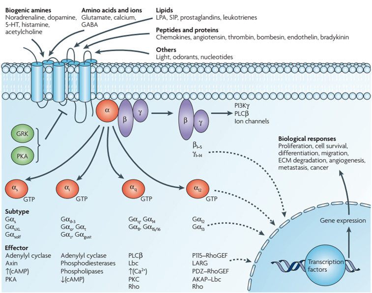
Gs, Gi and Gq route the same receptor architecture into different second messengers
| G protein | Primary effector | Second messenger | Typical consequence | High-yield memory |
|---|---|---|---|---|
| Gs | ↑ Adenylyl cyclase | ↑ cAMP | PKA activation | S = stimulates cAMP |
| Gi | ↓ Adenylyl cyclase | ↓ cAMP | less PKA signaling | I = inhibits cAMP |
| Gq | PLCβ | IP₃ + DAG + Ca²⁺ | PKC activation; ER Ca²⁺ release | Q → PLC → PIP₂ cleavage |
Cells actively shut signaling down
Desensitization means continued ligand exposure produces a smaller response. Receptors may be temporarily internalized and recycled, or sent to lysosomes for down-regulation.
Do not confuse G-protein signaling with receptor tyrosine kinase signaling

| Feature | GPCR | RTK |
|---|---|---|
| Membrane architecture | 7-pass receptor | single-pass receptor |
| Immediate transducer | heterotrimeric G protein | receptor kinase + phosphotyrosine docking proteins |
| Classic messengers | cAMP, IP₃, DAG, Ca²⁺ | Ras/MAPK, PI3K/Akt, PLCγ |
| Typical biological themes | rapid physiology, sensory and hormonal responses | growth, survival, differentiation and metabolism |
IMAT pattern: GPCR activation is GDP → GTP on Gα; Gs raises cAMP, Gi lowers cAMP, Gq raises IP₃/DAG/Ca²⁺. RTKs instead dimerize and autophosphorylate tyrosines.
9.6 Methods & Tools in Biology
Microscopy
- Light Microscope: Resolution ~200nm. Living cells visible. Stains (H&E) needed for contrast.
- Fluorescence Microscope: Uses specific wavelengths to excite fluorophores (e.g., GFP, DAPI). Localization of specific proteins.
- Transmission Electron Microscope (TEM): Resolution ~0.1nm. Electrons pass through specimen. 2D internal ultrastructure.
- Scanning Electron Microscope (SEM): Electrons scatter off surface. 3D topography.
Cell Fractionation
Separation of organelles by centrifugation speed.
- 600 g: Nuclei, Whole cells.
- 15,000 g: Mitochondria, Lysosomes, Peroxisomes.
- 100,000 g: Microsomes (ER/Golgi fragments).
- 300,000 g: Ribosomes, Viruses, Macromolecules.
9.7 The Three Questions Behind Most Cell-Biology Problems
Cell behavior emerges from three systems working together: membrane transport controls composition, the cytoskeleton organizes movement and shape, and signaling networks convert extracellular information into biochemical responses. These are tested as linked mechanisms rather than isolated definitions.
Cytoskeleton — structure predicts function
Actin treadmilling
ATP-actin is added preferentially at the plus end; turnover supports lamellipodia, microvilli, cytokinesis and cortical remodeling.
Dynamic instability
Microtubules alternate between growth and catastrophe. GTP-tubulin stabilizes a growing plus end; loss of the GTP cap favors shrinkage.
Intermediate filaments
They lack motor-protein tracks and are built for mechanical resilience. Tissue-specific IFs are useful histological markers.
Motor proteins
Kinesin usually moves toward microtubule plus ends; cytoplasmic dynein usually moves toward minus ends/centrosome.
Endocytosis, lysosomes and autophagy
Cell-mechanics pattern: actin drives cortex/motility, microtubules organize long-range transport and the spindle, intermediate filaments provide tensile resilience, and endosomes decide whether membrane proteins are recycled or degraded.
Viruses are obligate intracellular parasites. They contain genetic information and evolve, but lack ribosomes, independent metabolism and the complete machinery needed for reproduction. Therefore they are not cells and lie outside classical cell theory.

Viral structure: A genome is enclosed by a protein capsid; some viruses also possess a host-derived lipid envelope containing viral attachment proteins.
| Feature | Cell | Virus |
|---|---|---|
| Plasma membrane | Essential cellular boundary | Some viruses have envelopes, but this is not a cellular plasma membrane |
| Ribosomes | Present | Absent |
| Independent metabolism | Present | Absent |
| Replication | Cell division | Assembly using host-cell machinery |
Viruses
Apply this section immediately with official IMAT past-paper questions. Select an answer to reveal the full worked explanation.
Which of the following is absent from the genome of a virus that utilizes DNA as its primary molecule?
The decisive statement is Uracil
This question tests Viruses. Apply the definitions, structural relationships, and cause-and-effect rules in the section immediately above; the remaining choices conflict with at least one of those conditions.
Test every condition in the stem independently. Reject an option as soon as one statement contradicts the biological rules established in this lesson.
Which of the following types of nucleic acid is/are found in viruses?
1 single-stranded DNA
2 double-stranded DNA
3 RNA
The decisive statement is 1, 2 and 3
This question tests Viruses. Apply the definitions, structural relationships, and cause-and-effect rules in the section immediately above; the remaining choices conflict with at least one of those conditions.
Test every condition in the stem independently. Reject an option as soon as one statement contradicts the biological rules established in this lesson.
Which of the following is/are features of HIV?
1 It has a capsid.
2 It contains DNA.
3 It has ribosomes.
The decisive statement is 1 only
This question tests Viruses. Apply the definitions, structural relationships, and cause-and-effect rules in the section immediately above; the remaining choices conflict with at least one of those conditions.
Test every condition in the stem independently. Reject an option as soon as one statement contradicts the biological rules established in this lesson.
Which of the following molecules are associated with the Human Immunodeficiency Virus (HIV)?
1. DNA
2. RNA
3. Phospholipids
4. Reverse transcriptase
The decisive statement is 2, 3 and 4 only
This question tests Viruses. Apply the definitions, structural relationships, and cause-and-effect rules in the section immediately above; the remaining choices conflict with at least one of those conditions.
Test every condition in the stem independently. Reject an option as soon as one statement contradicts the biological rules established in this lesson.
The original 31 questions are retained and expanded with additional advanced mechanism questions. This revised lesson now also integrates organelle-atlas content such as proteasome vs lysosome, cell fractionation, specialized cells and endosymbiotic evidence. All questions are displayed in a single column.